Title: Ruoss, G.
Source text: The Medical and Surgical History of the War of the Rebellion. (1861-65.), Part 3, Volume 2 (Washington, DC: Government Printing Office, 1883), 183-184.
Civil War Washington ID: med.d2e8312
TEI/XML: med.d2e8312.xml
CASE 373.—Private G. Ruoss, Co. G, 7th New York, aged 27 years, was wounded near Petersburg, March 31, 1865, and admitted to the field hospital of the 1st division, Second Corps, where Surgeon F. M. Hammond, 126th New York, noted: "Shot fracture of right thigh." On the day following the injury the wounded man was sent to the Depot Hospital at City Point, and several days afterwards he was conveyed to Washington, where he entered Campbell Hospital, and subsequently Stanton Hospital. Surgeon R. B. Bontecou, U. S. V., reported his admission to Harewood Hospital September 12th, and his condition as follows: "The patient was suffering from great deformity of the right limb, the result of a shot wound of the upper third of the thigh, fracturing the femur. When admitted he had so far recovered as to be able to sit up, and his constitutional state was tolerably good. The fractured parts had firmly united but with great deformity; the wound of entrance had healed, but there were still some discharges from sinuses in the thigh, and small fragments of necrosed bones were daily removed. The prospects of usefulness of the limb are unfavorable; otherwise the patient is doing tolerably well." At the closing of Harewood Hospital, May 1, 1866, the patient was transferred to the Post Hospital at Washington, where he was operated on by Assistant Surgeon W. Thomson, U. S. A., who described the injury and operation as follows: "The wound was caused by a minié ball, which entered on the anterior and outer aspect of the thigh about three inches below the great trochanter, passed inward and a little downward, comminuting portions of the upper and middle thirds of the femur, and escaped posteriorly near the middle of the gluteal fold. On June 8th, the patient was etherized and a triangular incision, three by three and a half inches, was made on the upper and outer side of the thigh, and several pieces of bone were removed. On September 30th the wound had almost healed. Three sinuses, evidently leading to necrosed bone, however, still existed, two of which were located on the upper and one on the lower surface. The patient's health was not very good, having suffered from several attacks of diarrhœa, and a severe attack of erysipelas which commenced near the wound and soon spread over the entire surface of the leg and foot. On December 31st, the sinuses were still open and discharging, and there was great deformity, with about five inches shortening, and almost complete anchylosis of the knee joint." Assistant Surgeon J. Brooke, U. S. A., who took charge of the patient in November, 1867, reported the termination of the case: "I found the limb in much the same condition as described by Dr. Thomson, except that a collection of pus, which had formed on the inner aspect of the thigh just above the knee, had been opened and a sinus found to connect with the seat of the fracture. This sinus, as well as those already described by Dr. Thomson, remained open, and the patient continued extremely feeble, became greatly emaciated, and suffered much from diarrhœa and frequent attacks of almost total loss of appetite. These symptoms continued until his death, which occurred on June 27, 1868. At the autopsy the liver was found to be enormously enlarged, weighing ten pounds and ten ounces, and the right lung contained a small mass of calcareous matter." The injured femur, with the os innominatum and patella attached, and portions of the tibia and fibula were contributed to the Museum by Dr. Brooke, and constitute specimen 5450 of the Surgical Section. The femur is imperfectly united, with great displacement and a deposit of foliaceous callus, and shows that extensive periostitis has taken place, and the patella and upper portions of tibia and fibula also show similar pathological changes. The photograph represented in the wood-cut (FIG. 140) was obtained at the Harewood Hospital, and contributed by Surgeon Bontecou. Other photographs of the patient, taken at the same hospital and at the Army Medical Museum, are represented by No. 40, Vol. 8, of Photo's of Surgical Cases, S. G. O.; Card Photo's, Vol. 2, p. 21, and Vol. 3, p. 27, and Surgical Photograph Series, Nos. 139, 178, 179, and 292.
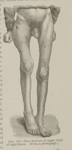 FIG. 140.—Shot fracture of upper third of right femur.
[From a photograph.]
FIG. 140.—Shot fracture of upper third of right femur.
[From a photograph.]