Title: Gardner, G. W.
Source text: The Medical and Surgical History of the War of the Rebellion. (1861-65.), Part 3, Volume 2 (Washington, DC: Government Printing Office, 1883), 18.
Civil War Washington ID: med.d2e816
TEI/XML: med.d2e816.xml
CASE 27.—Sergeant G. W. Gardner, Co. A, 12th Illinois Cavalry, aged 29 years, was wounded at Mitchell's Ford, on the Rappahannock, October 11, 1863. Surgeon S. B. Wylie Mitchell, 8th Pennsylvania Cavalry, reported that he was struck by a conoidal carbine ball, which entered four inches above the right knee, passed through the inner hamstring muscles and the adductor magnus, outward and downward, and lodged under the integument on the outer side of the thigh. Upon extracting the ball through an incision there was a profuse hæmorrhage, which ceased spontaneously. The patient was sent by railway to Washington, and entered Emory Hospital on October 13th. Acting Assistant Surgeon J. Walsh reported that "he was feeble and pallid; no pulsation could be detected in the tibial arteries of the injured limb. There was loss of sensibility and slight discoloration of the skin of the right foot. The leg was packed in raw cotton, and the temperature was kept up by bottles of hot water. Frictions with a stimulating liniment were occasionally employed. After October 18th, the leg was daily immersed in a bath of oxygen gas. On October 25th, two bits of blue cloth and a small piece of white cotton cloth were extracted from the wound. The foot was decidedly gangrenous, and gangrene began to advance rapidly up the leg." At this time a sketch of the appearances of the limb was made, under Surgeon J. H. Brinton's direction, by Hospital Steward E. Stauch. This drawing was elaborated, after the lamented death of Mr. Stauch, by Hospital Steward Schultze, and has been reproduced by chromolithography in the plate opposite (PLATE I, 28). On October 29th, Acting Assistant Surgeon W. H. Ensign amputated the limb at the lower third of the thigh. On examination of the amputated member it was found that the artery had been completely divided by the ball near the point at which it passes through the opening in the great adductor. The divided extremities of the artery were occluded by dark grumous clots. After the operation the patient was attacked by diarrhœa, and, on December 5, 1863, the case terminated fatally. A wet preparation of the lower half of the right femur, with a portion of the soft tissues, including the popliteal artery and vein, was contributed to the Army Medical Museum by Acting Assistant Surgeon J. Walsh (Cat. Surg. Sect., 1866, p. 521), and is represented in the adjacent wood-cut (FIG. 3).
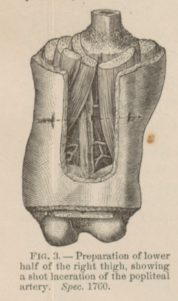 FIG. 3.—Preparation of lower half of the right thigh, showing a shot laceration of the popliteal artery. Spec. 1760.
PLATE XXVIII, facing p. 18. GANGRENE FOLLOWING A SHOT LACERATION OF THE FEMORAL ARTERY. (Chromolithograph.)