Title: Kagrice, J.
Source text: The Medical and Surgical History of the War of the Rebellion. (1861-65.), Part 3, Volume 2 (Washington, DC: Government Printing Office, 1883), 171.
Civil War Washington ID: med.d2e7451
TEI/XML: med.d2e7451.xml
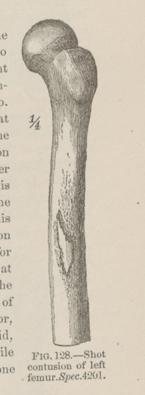
CASE 341.—Private J. Kagrice, Co. F, 198th Pennsylvania, aged 30 years, was wounded during the engagement at White Oak Road, March 31, 1865. Surgeon W. R. DeWitt, U. S. V., recorded his admission to the field hospital of the 1st division, Fifth Corps, with "shot wound of left thigh," and his transfer to City Point on the following day. Several days afterwards the wounded man was transferred to Douglas Hospital, Washington, whence Assistant Surgeon W. F. Norris, U. S. A., contributed the specimen (Cat. Surg. Sect., 1866, p. 258, Spec. 4201), with the following history: "The injury was a severe gunshot contusion of the left femur at its middle third. The patient was in good health and spirits at the time of his admission and doing well. The wound was carefully examined, but the ball could not be traced. On April 23d, a large abscess was opened on the anterior aspect of the thigh near the trochanter. On the 29th, another similar incision was required in order to give free exit to pus. The patient, however, continued pretty well until May 5th, when he had a chill. This recurred on May 7th and 8th, and on the 9th, 12th, 13th, and 14th, he had two chills a day. The thigh became painful, the discharge thin and fetid. On May 16th, the patient became delirious, had another chill, and his tongue became swollen and inflamed. He died on the following day. At the autopsy no pleurisy or effusion was found in either thoracic cavity. The lungs, liver, spleen, kidneys, and brain were carefully examined for pyæmic abscesses, but appeared healthy. On examining the femur it was found that the ball had struck it at its middle third, contused the bone without fracturing, and, being deflected from its course, had lodged in the hollow above the acetabulum. The hip joint was healthy and uninjured. A longitudinal section was made of the femur, which exhibited well marked gangrenous osteomyelitis,¹ the medulla being of a dirty greenish color, dry, pulverulent, and excessively fetid. The interspaces between the cancelli contained a dark greenish liquid, a quantity of which escaped in sawing the bone, and was also extremely offensive. The bone where the missile had struck had become necrosed and nearly separated, and around this partial exfoliation a ring of new bone had formed." A portion of the injured femur is illustrated in the adjacent wood-cut (FIG. 128).
¹ Dr. J. A. LIDELL in his excellent paper on Contusion and Contused Wounds of Bone, with an Account of Thirteen Cases, in Am. Jour. Med. Sci., 1865, Vol. L, p. 17, gives the following as the principal pathological effects of contusions of bone: 1. Ecchymosis of the osseous tissue; 2. Ecchymosis of the medullary tissue; 3. Osteomyelitis of a simple character; 4. Necrotic osteitis; 5. Suppurative osteomyelitis; and 6. Gangrenous osteomyelitis.
