Title: Woodworth, Henry
Source text: The Medical and Surgical History of the War of the Rebellion. (1861-65.), Part 3, Volume 2 (Washington, DC: Government Printing Office, 1883), 118.
Civil War Washington ID: med.d2e6034
TEI/XML: med.d2e6034.xml
CASE 270.—Private Henry Woodworth, Co. A, 4th Vermont, aged 18 years, was wounded at the battle of Spottsylvania Court House, on May 11, 1864, by a conoidal musket ball, which entered the left thigh, just below the trochanter major, passed inward and forward, grooving the femur anteriorly at the level of the lesser trochanter, and lodging under the sartorius muscle. The patient was conveyed to the field hospital of the 2d division of the Sixth Corps, where the ball was removed through an incision at the edge of the sartorius. A week subsequently, he was placed on one of the trains for the Rappahannock, and was transferred from Fredericksburg to Washington, where, on May 25th, he was admitted to Harewood Hospital. His condition on admission was very unpromising; his pulse was quick and feeble; he was anæmic, and without appetite. He was placed upon a tonic regimen, but he did not improve. The wound discharged profusely; there was much pain in the joint, pain aggravated by the slightest movement, and pus had burrowed in every direction about the articulation. Surgeon R. B. Bontecou, U. S. V., in charge of Harewood Hospital, decided that an excision of the head of the femur offered the only possible chance of saving life, and, on July 1st, the patient having been anæsthetized by sulphuric ether, Dr. Bontecou proceeded to perform the operation. A curved incision, with its concavity forward, embracing the trochanter, readily exposed the joint. The muscular attachments were divided, and the head was easily disarticulated, the joint being disorganized and the round ligament destroyed. The continuity of the bone being uninterrupted, the upper extremity was readily turned out and sawn just below the point of impact of the ball. On examination of the portion of bone removed, it was found that much of the head had been absorbed, and that the remainder was carious. The specimen is represented in the accompanying wood-cut (FIG. 75). The neck and trochanters are covered with traces of the effects of periostitis. The cotyloid cavity was ulcerated. The wound was drawn together by adhesive strips, and the limb was dressed in a fracture apparatus with moderate extension. Every means of supporting the patient's strength was adopted, but he did not rally from the operation, and, sinking gradually, expired on July 2, 1864.
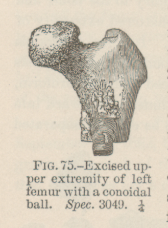 FIG. 75.—Excised upper extremity of left femur with a conoidal
ball. Spec. 3049. ¼
FIG. 75.—Excised upper extremity of left femur with a conoidal
ball. Spec. 3049. ¼