Title: M——, D. H.
Source text: The Medical and Surgical History of the War of the Rebellion. (1861-65.), Part 2, Volume 2 (Washington, DC: Government Printing Office, 1876), 67, 134, 163.
Civil War Washington ID: med.d2e31545
TEI/XML: med.d2e31545.xml
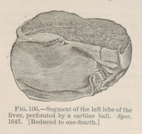
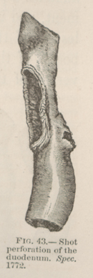
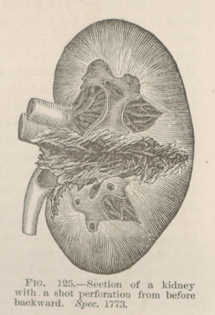
CASE 452.—Corporal D. H. M——, Co. H, 6th Pennsylvania Cavalry, in a cavalry engagement near Brandy Station, August 1, 1863, was shot through the body, the ball entering the right flank and escaping from the left of the epigastric region. There is no account of the symptoms observed at the field hospital station, which was crowded with wounded. The name, military description, and entry, "gunshot wound of the abdomen, simple dressing," constitute the only field record. On August 2nd, the corporal was transferred by rail to Washington, a distance of forty miles. He barely survived the transit, and expired, in an ambulance wagon on the way to Douglas Hospital. The dressings and clothing were saturated with blood, and there had evidently been very profuse bleeding after he was moved from the car to the wagon. At the autopsy, made by Medical Cadet Edward D. Mitchell, it was inferred, from the size of the wounds of entrance and exit, that they were inflicted by a carbine ball. The notes of the autopsy describe a very erratic course of the projectile; but if the position of the entrance and exit wounds is correctly indicated, it must be inferred that the ball entered the right hypochondrium at the edge of the twelfth rib, scraping oft its outer lamina about two and one-half inches from its free extremity, and passed inward and downward, penetrating the right kidney , and was then deflected by the vertebral column, and passed upward through the duodenum, the posterior and anterior walls of the stomach, the left lobe of the liver near the umbilical fissure (FIG. 106), and emerged two inches to the left and below the end of the ensiform cartilage; or its course may have been the reverse of that described. The intestines were inflated. There was a great quantity of blood in the peritoneal cavity. The emulgent vessels had bled with especial freedom. The perforation of the liver is represented in the wood-cut (FIG. 106). The wound of the duodenum is figured on page 67, and there will be a drawing of the perforation of the kidney in the subsection on wounds of that organ.


