Title: Hardy, Francis M.
Source text: The Medical and Surgical History of the War of the Rebellion. (1861-65.), Part 2, Volume 2 (Washington, DC: Government Printing Office, 1876), 950.
Civil War Washington ID: med.d2e30854
TEI/XML: med.d2e30854.xml
CASE 1897.—Private Francis M. Hardy, Co. B, 30th North Carolina, aged 22 years, was wounded near Fort Stevens, July 12, 1864, captured, and admitted to Lincoln Hospital, from the Defences of Washington, on the 14th. Assistant Surgeon W. Lindsly, U. S. A., reported: "Gunshot wound; a minié ball entering and shattering left ulna, lower third, passing through and entering right radius, middle third, shattering that bone and lodging." Acting Assistant Surgeon F. L. Leavitt described the case as follows: "Patient was wounded while lying behind the corner of a frame dwelling house, and in the act of firing, by a minié ball, which he states first passed through the angle of the house. Resection of both injured bones was performed on the field the same day, by Surgeon G. W. Briggs, 30th North Carolina, the arm placed on straight splints, and in this condition the patient was received in the hospital, suffering a good deal from the tightness of the bandages, the parts swelling considerably. Applied a loose water dressing merely sufficient to keep the bones in position. Signs of abscess at edge of right biceps; the exploring needle revealed pus, and the probe the presence of a foreign body which I removed, and it proved to be the bullet very much battered and irregular. 23th, doing well on iron and quinine. 29th, erysipelatous condition of left forearm; right upper extremity looks healthy. August 2d, left forearm entirely healed; right heals slowly. 8th, gangrene started in the wound of incision where the ball was extracted. 11th, arrested by bromine and stimulating poultices. 12th, patient delirous and very feverish; much prostrated and emaciated from the effects of the diarrhœa; gangrene again commenced and attacked the left forearm; bromine used with no success; brachial artery exposed for the space of two inches in right arm; a tourniquet was placed high up in the axilla ready for use if required. 13th, secondary hæmorrhage occurred this morning, about eight o'clock, from the right brachial artery, but was promptly checked and the artery tied. Afternoon, secondary hæmorrhage took place about two o'clock, in the other arm, from one of the muscular branches of the radial artery; the persulphate of iron arrested the bleeding. Evening, brachial artery sloughed through at point of ligation and a severe hæmorrhage followed; the blood being in a defibrinated condition all efforts at forming a clot failed. 17th, hæmorrhage from left ulnar artery occurred this morning, but was arrested by pressure; patient very much weakened; comatose. Died." Dr. Leavitt does not mention a hæmorrhage from the brachial, and the ligation of that artery, in his report of the case to the Surgeon General. In an account published in the Medical and Surgical Reporter, Vol. XIII, for 1865-6, p. 299, he states that the patient died on the 14th. At two o'clock on the afternoon of the 17th, Acting Assistant Surgeon H. M. Dean made an autopsy, and furnished the following notes: "Body considerably emaciated; rigor mortis well marked; height, five feet six inches. He has three sores which have evidently been the seat of phagedæna, viz: one on the inner surface of the left forearm; one on the outer aspect of the right forearm; and one on inner aspect of right arm just above the elbow; the former was in a state of gangrene. The lower half of the left ulna and a portion of the middle third of the right radius had been resected, the brachial artery stretched across the ulcer in the right arm having been entirely isolated from the surrounding tissues for a distance of two and a half inches. Lower lobe of right lung in a state of gray hepatization, with the exception of the upper portion of its free margin; posterior portion of the upper lobe also in the third stage of pneumonia, the rest of the right lung was normal; posterior portion of lower lobe of left lung in a state of gray hepatization, the rest of the lung was normal; right lung weighed twenty-three ounces, left thirteen ounces; pericardium normal; spleen normal, weight nine and a half ounces; left lobe of liver somewhat longer than usual; the right lobe was in the shape of a sugar-loaf; parenchyma normal, weight fifty-seven and one-half ounces; brain healthy, weight fifty-one and one-half ounces." The specimens, represented in the accompanying wood-cuts, were forwarded to the Museum by Dr. Dean. The first (FIG. 688) consists of the bones of the right forearm with the middle third of the radius excised; no reparative action exists at the extremities. The second (FIG. 689) consists of the bones of the left forearm from which the lower half of the ulna has been excised; the extremity of the bone shows a slight ring of necrosis and no attempt whatever at repair.
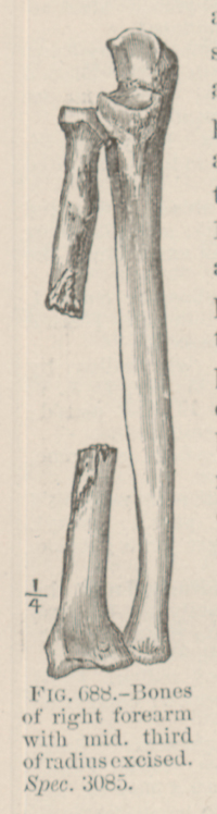 FIG. 688.—Bones of right forearm with mid. third of radius excised. Spec. 3085.
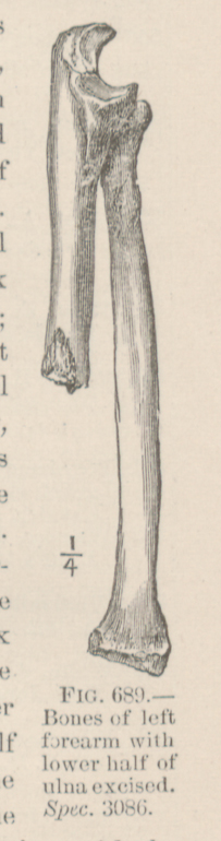 FIG. 689.—Bones of left forearm with lower half of ulna excised. Spec. 3086.