Title: Singleton, D.
Source text: The Medical and Surgical History of the War of the Rebellion. (1861-65.), Part 2, Volume 2 (Washington, DC: Government Printing Office, 1876), 574.
Civil War Washington ID: med.d2e30135
TEI/XML: med.d2e30135.xml
CASE 1556.—Private D. Singleton, Co. F, 140th Pennsylvania, aged 30 years, was wounded at Spottsylvania, May 12, 1864, and was treated in a Second Corps field hospital, and thence sent to Washington, entering Carver Hospital on May 16th. He was operated on by Surgeon O. A. Judson, U. S. V., who reported as follows: "Gunshot wound of shoulder; conoidal ball lodged in head of humerus, splitting the head and detaching splinter. On May 27th, a straight incision was made through the deltoid, and the head and two inches of the shaft of the humerus was removed by a chain saw. Result favorable, the wound filling with healthy granulations." The patient was transferred, and admitted to hospital at Pittsburg on November 16, 1864, and on June 11, 1865, was discharged the service and pensioned. Examiner D. Stanton, of New Brighton, Pennsylvania, May 31, 1868, reported: "Minié ball lodged in the head of the humerus. The head and about two inches of the shaft excised May 20, 1864. There is now a space of about two and a half inches between the upper end of the shaft and the glenoid cavity. There is still necrosis of the bone. There is very slight voluntary motion of the arm, and limited use of the forearm and hand." On June 23, 1866, J. Wilson Wishart, late Surgeon 149th Pennsylvania, forwarded to the Army Medical Museum a photograph exhibiting the appearance of the injured limb at that time. The arm is supported at the highest point to which he could raise the elbow by voluntary efforts. This photograph is represented by Figure 3 in PLATE XVIII. Dr. Wishart states: "During the present month Mr. S. called upon me at my office, and upon examination I found a slight discharge from the point indicated in the photograph by the dark spot at the junction of the upper and middle thirds of the cicatrix. Upon introducing a probe I discovered diseased bone, which, at his request, I removed on Thursday the 21st instant, through an incision in the upper part of the cicatrix. It proved to be a portion of the sawed surface of the humerus, which, with pieces previously discharged, completed about two-thirds of the circumference of the bone. A probe, introduced to the point whence the bone was extracted, entered a canal some two inches in depth, extending downward and to the inside of the humerus. Careful and repeated examination by myself and another surgeon failed to detect any communication with the humerus, or any more diseased bone. I am in hopes now that the wound will entirely close." In a certificate for increase of pension, January 16, 1867, Examiner Stanton reported: "There is now false joint, there being a space of two and a half inches between the upper end of the shaft and the glenoid cavity. The slight use of the hand is more than counterbalanced by the pain and soreness caused by its use. Important tendons were severed by the ball or by the knife of the surgeon." In September, 1873, a Board, convened at Pittsburg, and composed of Drs. A. G. McCandless, J. W. Wishart, and W. J. Elmore, reported: "The arm is useless for purposes of manual labor." The pensioner was paid June 4, 1874.
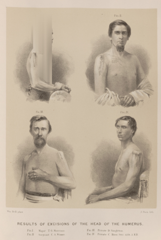 PLATE XVIII __ RESULTS OF EXCISIONS OF THE HEAD OF THE HUMERUS. FIG. III. Private D. Singleton.