Title: Donnelly, Reuben
Source text: The Medical and Surgical History of the War of the Rebellion. (1861-65.), Part 3, Volume 2 (Washington, DC: Government Printing Office, 1883), 721.
Civil War Washington ID: med.d2e29169
TEI/XML: med.d2e29169.xml
CASE 1067.—Private Reuben Donnelly, Co. A, 25th Ohio, was wounded at Bull Run, August 30, 1862. Acting Assistant Surgeon Thomas C. Barker reported: "He entered King Street Hospital, Alexandria, on September 3d, suffering with a wound of the right thigh. A round ball entered the front part of the thigh just above the patella and passed directly through the limb from front to rear, dividing the condyles and splitting the bone up the shaft several inches. On September 5th Surgeon J. E. Summers, U. S. A., amputated the thigh at the junction of the lower and middle thirds by the antero-posterior flap operation. The case progressed favorably. Union had taken place throughout the whole of the flaps, with the exception of one point, about one-eighth of an inch in diameter, over the end of the bone, which lay at a depth of three-fourths of an inch below the orifice of the aperture. Patient's health, appetite, and general vigor had been good up to the night of October 3d, when, during a cold rain, he was attacked with a severe chill. The next day, in the afternoon, he had another chill, and although tonics and stimulants were sedulously used with him, he sank in general vigor and his appetite failed. Absorption of the new granulations took place around the aperture which had remained ununited, and the opening increased to nearly an inch in diameter, the integuments retracting and leaving the end of the bone nearly on a level with the external surface. Suppuration occurred in the medullary canal of the bone. Emaciation was extreme; debility was rapidly increasing, with indisposition and incapacity of taking and retaining food, or even stimulants to any appreciable amount; and the patient gradually sank, and died of exhaustion and debility October 20, 1862." The specimen, consisting of the lowest third of the right femur, perforated by a round ball directly through the centre of the shaft, just below the upper margin of the patella, was forwarded to the Museum by Dr. Barker, and is represented in the wood-cuts (FIGS. 403, 404). The shaft is split longitudinally in its anterior surface for four inches, and posteriorly obliquely for the same distance. The condyles are also separated by a fissure.
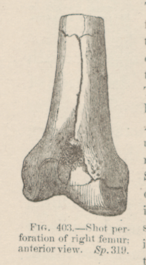 FIG. 403.—Shot perforation of right femur; anterior view.
Sp. 319.
FIG. 403.—Shot perforation of right femur; anterior view.
Sp. 319.
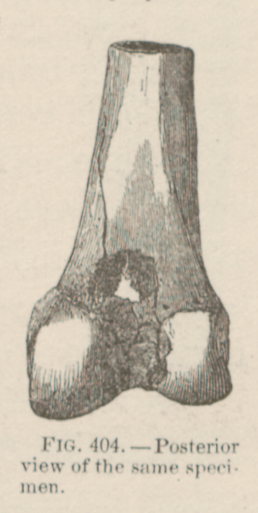 FIG. 404.—Posterior view of the same specimen.