Title: Mock, L.
Source text: The Medical and Surgical History of the War of the Rebellion. (1861-65.), Part 3, Volume 2 (Washington, DC: Government Printing Office, 1883), 529, 712-713.
Civil War Washington ID: med.d2e20548
TEI/XML: med.d2e20548.xml
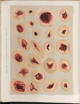
CASE 777.—Private L. Mock, Co. I, 119th Pennsylvania, aged 22 years, was wounded in the left foot, at Rappahannock Station, November 7, 1863, by a musket ball, which entered about one and a half inches below the internal malleolus, passing upward and outward, making its exit anterior and under the external malleolus and opening the ankle joint in its passage through the parts. He entered Armory Square Hospital at Washington two days after receiving the injury. On December 6th, Surgeon D. W. Bliss, U. S. V., amputated the leg above the ankle by flap operation, the patient being under ether, to which a little chloroform was added towards the last. Three vessels were ligated, the loss of blood being small. The patient reacted well from the anæsthetic. At the time of the operation his constitutional condition was tolerably good and the tissues of the wounded parts were in a healthy state. The stump grew painful, but its appearance was good. Cold-water dressing was used and nourishing diet with stimulants were administered. On December 9th the patient had three chills, followed by profuse perspiration. On the following day there was no chill, the stump still looked well, and the patient suffered no pain. Quinine and opium were now prescribed in addition to the former treatment. By December 18th there was well-developed pyæmia, from the effects of which the patient died December 21, 1863. After death yellowness, to an extreme degree, came on, and, on inspection, the shoulder and elbow were found to contain pus, the shoulder having a large quantity and the other joints being believed to be in a like condition. The tarsal bones of the amputated limb, contributed with the history by the operator, constitute specimen 1903 of the Surgical Section of the Museum. The specimen exhibits no perceptible attempt at repair, and shows that the ball passed through the calcaneum, which is necrosed, and grazed the astragalus, opening the ankle joint. The appearance of the entrance and exit wounds is shown in PLATE XXXIX, FIGURE 2.
