Title: Heakin, J.
Source text: The Medical and Surgical History of the War of the Rebellion. (1861-65.), Part 3, Volume 2 (Washington, DC: Government Printing Office, 1883), 521.
Civil War Washington ID: med.d2e20161
TEI/XML: med.d2e20161.xml
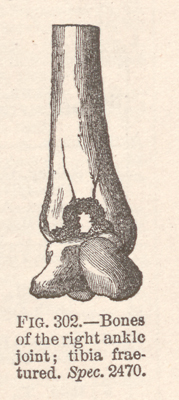
CASE 768.—Private J. Heakin, Co. D, 6th U. S. Cavalry, aged 24 years, was wounded in the right ankle, at the battle of Old Church, May 31, 1864. He was admitted to Stanton Hospital, Washington, four days afterwards, where amputation was performed by Surgeon J. A. Lidell, U. S. V., who made the following report:¹ "The wound was inflicted by a minié ball, which entered anteriorly, passed backward and inward, apparently going close to the posterior tibial artery and escaping behind the lower end of the tibia. The ankle joint was involved. The parts became swollen, red, and tender. During the night of June 7th the patient had secondary hæmorrhage from the wound, losing about a pint of blood, bright red in color. On the following day the leg was amputated at the place of election by double flap method, the anterior flap being shorter than the posterior, and the tibia being divided after the procedure of Sanson. Sulphuric ether constituted the anæsthetic. The loss of blood was trifling during the operation and the patient's general condition at the time was favorable, there being no constitutional disturbance worth mentioning. The shock of the operation was little and passed away quickly; reaction moderate. The patient died of pyæmia June 21, 1864. The examination of the injured member showed the lower end of the tibia to be badly comminuted into the ankle joint, which was filled with pus. The posterior tibial artery was grazed by the bullet and some very small fragments of bone had been driven into it. The hæmorrhage occurred on the detaching of these fragments by suppuration, together with the separation of the bruised tissue belonging to the wall of the artery. The astragalus was uninjured." The latter bone and the lower portion of the amputated tibia (Spec. 2470) were contributed to the Museum by the operator, and are represented in the wood-cut (FIG. 302).
¹ LIDELL (J. A.), Secondary Hæmorrhage from Posterior Tibial Artery, etc., in U. S. Sanitary Commission Memoirs, New York, 1870, Vol. I, p. 23.
