Title: Esworthy, W. N.
Source text: The Medical and Surgical History of the War of the Rebellion. (1861-65.), Part 3, Volume 2 (Washington, DC: Government Printing Office, 1883), 454-455.
Civil War Washington ID: med.d2e18895
TEI/XML: med.d2e18895.xml
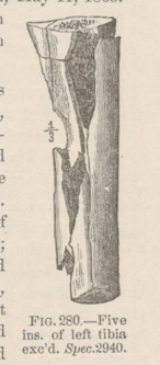
CASE 717.—Corporal W. N. Esworthy, Co. E, 1st Pennsylvania Cavalry, aged 24 years, was wounded in the left leg at White House Landing, June 21, 1864, and admitted to Carver Hospital, Washington, two days afterwards. Surgeon O. A. Judson, U. S. V., reported: "The missile, supposed to be a conoidal ball, entered the leg anteriorly about three inches below the knee joint, passed backward and outward, making its exit posteriorly and producing a compound comminuted fracture of the upper third of the tibia. Supporting treatment was adopted and simple dressings applied. Erysipelas attacked the wound on June 27th, but yielded readily to an application of solution of copperas. By July 1st no erysipelatous symptoms were present; constitutional state of patient good; wound secreting laudable pus in large quantity. On July 6th, the patient was anæsthetized and excision of about five inches of the upper third of the shaft of the tibia, by means of the chain saw, was performed by Acting Assistant Surgeon O. P. Sweet, making a straight incision over the crest of the tibia. The wound was then filled with scraped lint, cold-water dressings were applied, and stimulants given freely. The patient appeared to be doing quite well up to July 18th, the wound filling with healthy granulations and secreting laudable pus. For ten days previous, however, he had anorexia, and this morning he had a very severe chill. A recurrence of chills followed every morning and sometimes two or three times during the day, and these were followed by other pyæmic symptoms, the integuments assuming a deep icteric tinge; pulse rapid; slight cough; anorexia continuing. The treatment, decidedly stimulating and tonic, was continued. The symptoms continued in a more aggravated form and the patient steadily sank, the whole surface of his body being of a deep yellow color. He died on July 23, 1864. At the autopsy a large number of metastatic abscesses were found in both lungs; liver enlarged; spleen also enlarged, dark colored, and soft. A purulent offensive fluid mixed with lymph was shown in the left pleural cavity, and a large disintegrated clot in the femoral vein." The excised bone, contributed by Surgeon Judson and exhibiting superficial necrosis, is represented in the wood-cut (FIG. 280).
