Title: Rall, Henry
Source text: The Medical and Surgical History of the War of the Rebellion. (1861-65.), Part 3, Volume 2 (Washington, DC: Government Printing Office, 1883), 428-429.
Civil War Washington ID: med.d2e17100
TEI/XML: med.d2e17100.xml
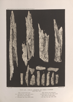
CASE 657.—Corporal H. Rall, Co. D, 15th New York Heavy Artillery, aged 22 years, was wounded at the battle of Old Church, May 30, 1864. Surgeon L. W. Read, U. S. V., reported his entrance into the field hospital of the 3d division, Fifth Corps, with "shot wound of left leg." Surgeon B. B. Wilson, U. S. V., in charge of Stanton Hospital, Washington, contributed the pathological specimen (No. 4337 of the Surgical Section, A. M. M.), with the following history: "This young man was admitted to this hospital on June 4th. He had been wounded by a minié ball, which passed across the spine of the tibia about four inches from the head of the bone, bruising it and denuding it of periosteum along the track of the ball. He was somewhat debilitated when admitted, but being young and of good constitution, his general condition was not unfavorable. He was treated with applications of cold water and ice dressings to the wounded limb, and stimulating and supporting constitutional remedies. During the month of June and the beginning of July the limb was highly inflamed, and there was profuse suppuration and some sloughing in the vicinity of the wound; with considerable sympathy of the general system as manifested by chills, great debility, loss of appetite, and general febrile action. In the month of July, deep-seated fluctuation having been observed in the course of the shaft of the tibia, the pus was evacuated by free incisions in the direction of the length of the limb, with great relief to the patient. The periosteum was found to be extensively separated and the shaft of the bone necrosed. During the months of September, October, November, and December his condition gradually improved as the process of formation of the involucrum and the separation of the necrosed portion went on. A number of cloacæ formed in the line of the incisions for liberating pus, through which the necrosed bone could be felt gradually becoming detached from the living portion. The limb was, during this time, for the most part treated with emollient poultices. About the first of January the upper part of the shaft of the tibia could be distinguished at the position of the original wound, and about March 1st, upon seizing it with a forceps the whole dead mass could be moved within the sheath of the investing new bone. The operation for the removal of necrosed bone was performed by Surgeon A. N. Dougherty, U. S. V., on March 14, 1865, by turning back the soft parts on each side from an incision through the centre of the cloacæ, and cutting away with the mallet and chisel sufficient of the new growth to permit the sequestrum to be lifted directly from its bed. It was found to consist of the entire shaft of the tibia from one epiphysis to the other, except small portions eroded by the absorbents. The after-treatment consisted of simple water dressings with slightly stimulating applications, and was unmarked with any noteworthy complication. On June 6, 1865, the patient was discharged on certificate of disability, being able to walk with ease and comfort, though the wound was not entirely healed. In July a photograph was taken, of which the adjoining wood-cut (FIG. 257) is a copy. He left the hospital in July and returned in the following month, asking to be employed under contract. Since that date he has been doing duty as chief nurse of one of the wards of this hospital, being in robust health, though his limb was not yet entirely healed." The specimen, consisting of a sequestrum nine inches long and eleven smaller pieces of necrosed bone, is shown somewhat reduced in FIG. 1 of PLATE LXXI, opposite page 428. Examiner T. F. Smith, of New York City, September 22, 1873, certified: "Shot fracture of left tibia, with union and great loss of bone substance, leaving a cicatrix over the anterior surface of the bone nine inches in length, red, unhealthy, and ulcerating," etc. The Brooklyn Examining Board reported, September 8, 1877: "We find an adherent, chronically inflamed cicatrix extending along the anterior face of the left tibia from below its head to within three inches of the ankle. There is tenderness on pressure. He requires the application of a bandage, and complains of pain in damp or cold weather. The usefulness of the limb is well nigh destroyed." The pensioner was paid March 4, 1880.
FIG. 257.—Appearance of limb fourteen months after injury. [From a photograph.]
