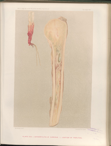
Title: Aseltyne, M. B.
Source text: The Medical and Surgical History of the War of the Rebellion. (1861-65.), Part 3, Volume 2 (Washington, DC: Government Printing Office, 1883), 405, 736 (Part 2, Volume 2).
Civil War Washington ID: med.d2e16821
TEI/XML: med.d2e16821.xml
CASE 649.—Private M. B. Aseltyne, Co. F, 10th Vermont, aged 22 years, was wounded in the left foot, at Mine Run, November 27, 1863, and entered the Third Division Hospital, Alexandria, one week afterwards. Acting Assistant Surgeon A. P. Crafts reported: "A conical ball entered the anterior surface of the foot, fractured the cuneiform bones, and was extracted through the orifice of entrance, on the field. At the time of the patient s admission there was much tumefaction and inflammation of the foot and leg, which at first seemed to yield to treatment. But, on December 10th, evidences of gangrene began to show themselves, and four days afterwards the leg was amputated at the knee joint by Surgeon E. Bentley, U. S. V. The operation was performed by the circular method, and by retaining the patella after disarticulating the joint. Chloroform constituted the anæsthetic. Simple dressings were used to the stump, and tonics and stimulants, including iron and quinine, and acetate of ammonia were given in the treatment. On December 20th, there were symptoms of pyæmia. The patient's countenance became sallow and anxious, a severe chill occurred, and hiccough set in, followed by loss of appetite and by profuse sweating. Death supervened on December 27, 1863. At the post-mortem examination the liver, kidneys, spleen, and intestines were found healthy, the lungs much discolored, and some effusion in the cavity of the chest. There was also great hypertrophy of the heart and some effusion within the pericardium." A section of the stump, made by Surgeon J. H. Brinton, U. S. V., showed the femur to be healthy, although denuded of periosteum for six inches above the joint. The synovial membrane on the crucial ligament was congested, and the cartilage of the femur was thinned and softened, the whole color being changed and absorption commencing. The ligature of the popliteal artery had partially sloughed, and the base of the long internal clot had come down and projected through the opening made by sloughing in the walls of the vessel. The artery, showing its condition as described, was contributed to the Museum by the operator. A wet preparation of it constitutes specimen 1989 of the Surgical Section, and a chromo-lithographic representation is shown on PLATE. XXII, opposite page 736 of the Second Surgical Volume.
