Title: Pool, John
Source text: The Medical and Surgical History of the War of the Rebellion. (1861-65.), Part 3, Volume 2 (Washington, DC: Government Printing Office, 1883), 306-307.
Civil War Washington ID: med.d2e13558
TEI/XML: med.d2e13558.xml
CASE 479.—Private John Pool, Co. H, 119th Pennsylvania, aged 23 years, was wounded by a conoidal ball, at Rappahannock Station, November 7, 1863. He was admitted into the Stanton Hospital, Washington, November 9th. Surgeon John A. Lidell,³ U. S. V., furnished the following history, with the specimen (No. 2229, Sect I), to the Army Medical Museum: "The bullet had entered the lateral and external part of the left thigh near the junction of the middle and upper thirds, and passing downward, inward, and somewhat forward, had fractured the femur at its middle. The missile appears to have been split, by contact with the bone, into two pieces, which made their escape through separate openings in the popliteal space. When admitted the knee was much swollen and hot; the patella floated some distance above the femoral condyles. He did not complain of pain, and his general condition was favorable. The wounded limb was propped up with long sand bags placed on either side of it, and moderate extension was applied by a weight hanging over the foot of the bed and attached to the foot and leg by strips of adhesive plaster. Counter extension was effected by elevating the foot of the bed. He was put upon supporting diet, and simple dressings applied. The inflammation and swelling at the knee gradually subsided, and, on December 1st, had entirely disappeared, the wounds in the popliteal space closed; the wound of entrance, however, was still open and discharging. On the 20th, there was a slight hæmorrhage of arterial blood from the wound of entrance, which was readily checked. On the 28th, the limb was placed in Hodgen's splint to facilitate the outflow of pus. January 8, 1864, the wound in the popliteal space reopened and the wounds, both of entrance and exit, discharged freely. 24th, pulse 110; he was daily becoming more feeble. On exploring the wound through the orifice of exit with Nélaton's probe detached fragments were found at the seat of fracture. The patient was etherized, and through an incision about four inches long, made in the back part of the thigh, six detached fragments of considerable size, the largest about two inches long by three-fourths of an inch wide, were extracted. The superior and inferior part of the fractured femur were found to be held in proper position by strong splints of provisional callus, which had been thrown out on the inner and outer sides of the bone. The finger could be readily passed between the broken extremities of the bone, both ends of which were necrosed but not yet detached. On the 25th, the thigh had swelled a good deal and was emphysematous, but, on the 28th, the emphysema had disappeared and the swelling was subsiding; the limb was again placed in Hodgen's splint. On the 31st, he was improving in every respect. On February 14th, he was doing finely; no night sweats; slept well; appetite good; pulse 80; discharge of pus moderate. 24th, two fragments of bone were extracted. March 1st, a diffuse inflammation, accompanied with redness and much swelling, attacked the thigh and spread rapidly through the limb; there was also great constitutional disturbance. After a time this inflammation subsided in a great measure but left him much weakened. About April 1st, another attack of diffuse inflammation brought him still lower. 18th, the whole limb was greatly swollen from the groin to the toes; a small slough on the instep separated; the knee joint was extended with effusion, the patella floating some distance above the femoral condyles. There was a profuse discharge of thin pus from the wound of operation in the back part of the thigh. He was much emaciated, pale, and weak; pulse frequent and feeble; tongue red and inclined to be dry; appetite capricious and poor, and he was subject to frequent attacks of diarrhœa. He was steadily failing, and, there being no hope of saving his life without amputation, he was placed under the influence of sulphuric ether, digital compression applied to the femoral artery, and the thigh amputated in the upper third, by the double flap method, by Surgeon J. A. Lidell, U. S. V. The femur was sawn off about one and a half inches below the trochanter minor. The soft parts of the thigh were so much diseased as not to admit the performance of the operation at any point below. But little blood was lost during the operation. There was a good deal of shock, but he reacted promptly afterward. In a short time the stump became sloughy; he gradually failed, and died of exhaustion April 26, 1864. On examining the amputated member, extensive burrowing of pus was found among the muscles of the thigh, and numerous small pieces of the bullet and fragments of bone sticking into the soft part around the seat of fracture. The ends of the broken femur were not in apposition, but separated from each other by the space which had formerly been occupied by the fragments of bone extracted by operation. Pretty firm union had, however, taken place by means of a bridge of new bone which arched over the chasm in front. On splitting the femur open lengthwise with a saw the marrow presented a coppery-red color, and there were abundant, deposits of new reddish colored osseous tissue both within the medullary canal, endostosis, and, external to the bone, periostosis, for a considerable distance above the fracture. In the marrow below the fracture there was a large-sized chocolate-colored spot, the result, apparently, of an old extravasation of blood. The substance of the marrow was decidedly tougher than normal. There was a considerable deposit of new osseous tissue lying between the periosteum and the bone. The periosteum itself was thicker and redder than natural in that locality, and from it these laminæ of new osseous tissue had been developed. The knee joint was swelled out with a straw-colored jelly-like substance; the synovial fringes were reddened, but the articular cartilage presented no abnormity." A drawing of the specimen, No. 2229, by Hospital Stewart E. Stauch, is copied in PLATE LXVI, opposite p. 306.
³ LIDELL (J. A.), On Secondary Traumatic Lesions of Bone, viz: Osteo-myelitis, Periostitis, Ostitis, Caries, and Necrosis, in U. S. Sanitary Commission Memoirs, 1870, Surgical Volume I, p. 414.
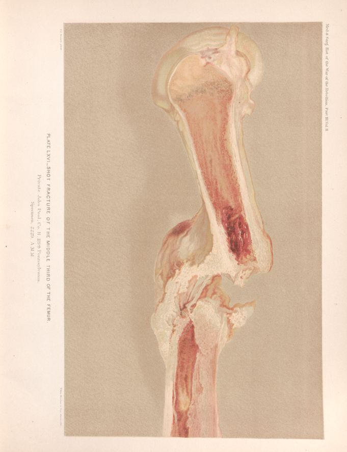 PLATE LXVI __ SHOT FRACTURE OF THE MIDDLE THIRD OF THE FEMUR. Private John Pool, Co. H, 119th Pennsylvania. Specimen, 2229, A. M. M.