Title: Sproul, John
Source text: The Medical and Surgical History of the War of the Rebellion. (1861-65.), Part 3, Volume 2 (Washington, DC: Government Printing Office, 1883), 225-227, 228-229.
Civil War Washington ID: med.d2e10923
TEI/XML: med.d2e10923.xml
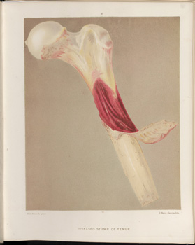
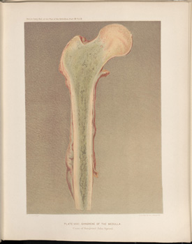
CASE 440.—Sergeant John Sproul, Co. C, 40th New York, aged 24 years, received at Kelly's Ford, November 7, 1863, a conoidal musket ball wound, causing a compound fracture of the left femur just above the knee. He was taken to the field hospital of the 1st division, Third Corps, Surgeon J. W. Lyman, U. S. V., in charge, and amputation of the thigh in the middle third, anterior and posterior flap method, was performed by Surgeon A. Campbell, 40th New York, on the day of injury. He was conveyed to Washington, and admitted into the Douglas Hospital on November 9th. His attending medical officer, Assistant Surgeon W. F. Norris, U. S. A., reports, November 16th: It was found impossible to support the heavy posterior muscular flap; the sutures sloughed out, and there was a great gaping surface discharging profusely, but normal in appearance. The patient was very pallid. The stump was thoroughly supported by adhesive straps, and the best nutrients, with stimulants, were administered. 24th, there was slight tenderness along the femoral artery and slight enlargement of the inguinal glands. There was no vigorous effort at repair in the stump, which remained pale and flabby, and his general condition became worse daily. December 4th, there were well-marked chills with fever in the evening, pulse 120 and feeble, respiration hurried, sweating, and nausea. These symptoms became hourly worse; his pulse became countless, his respiration sighing and very rapid, his face pinched and anxious; occasional vomits of a green bilous matter: he finally died, on December 7th, at 4 o'clock P. M., of pyæmia. Autopsy sixteen hours after death. Assistant Surgeon W. Thomson, U. S. A., records: "The most careful examination failed to find a trace of inflammation or coagulation in the blood-vessels. The soft tissues of the stump were perfectly normal. There were found in the thoracic cavity the usual traces of pyæmia. The lungs anteriorly were pale, posteriorly they were both coated over the lower lobes with recent yellow and soft ill-looking lymph. There was no considerable effusion or adhesions; no traces of a frank pleuritis, but of a local asthenic inflammation were found. Both lungs were congested, hardened, and dark in color posteriorly, and in the left there were several small yellowish-white spaces, apparently abscesses. * * The other organs, except the cerebro-spinal, were examined and found normal except the spleen, which was slightly hardened and congested. No trace of embolia was found in the lungs, nor was death caused by the secondary changes there produced. The destruction of pulmonary tissue had not progressed sufficiently. There must have been the absorption, by the veins most probably, from this medullary and cancellated portion of the femur, of a material soluble in the fluids of the blood, and produced, in the cadaveric changes that took place in the organic matter—dead, but confined in this cancellated bone. This caused the fatal toxicæmia which overpowers the nervous system, and may have also caused those local changes found in the posterior portions of both lungs. It seems strongly probable that it is due to blood poisoning, introduced by the veins, since all cases of pyæmia exhibit pathological changes in the lungs, where the venous blood becomes distributed to thin delicate structures before being depurated by exposure to the atmosphere. The case was about to be relinquished as incomprehensible, when it was proposed by Dr. Norris to saw the femur from its head to its extremity longitudinally, and thus expose its medullary canal. A small quantity of apparently healthy pus had been found between the periosteum and the shaft two inches from the sawn end, and there were a few osteophytes clinging to the bone at that point. When, however, the bone had been separated in its long diameter, its medullary canal presented the traces of pathological action. This canal and the cancellated structure extending past the trochanters to a point half way through the head were found filled with a yellowish-green substance, intolerably fetid, and resembling, more than anything else, the debris of hospital gangrene. The bone was sent at once to the Museum and portrayed in colors. No pus was found in this bone, and, under the microscope, nothing but debris. The connective tissue seemed to have perished en masse. In PLATE XXXII, opposite, the diseased stump of the femur is represented, and PLATE XXXI, opp. p. 228, exhibits the gangrenous condition of the medullary canal.

