Title: Chappel, Benjamin F.
Source text: Surgeon General Joseph K. Barnes, United States Army, The Medical and Surgical History of the War of the Rebellion. (1861–65.), Part 1, Volume 2 (Washington, D.C.: Government Printing Office, 1870), 124-125.
Civil War Washington ID: med.d1e8987
TEI/XML: med.d1e8987.xml
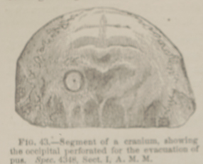
CHAPPEL, BENJAMIN F., Sergeant, Co. H, 8th New York Cavalry, aged 27 years, was wounded, before Petersburg, Virginia, April 1st, 1865, by a pistol ball which entered one inch above and one and a half inches to the left of the occipital protuberance and emerged just below it on the opposite side, denuding the bone of pericranium. He was admitted to the hospital of the 3d division, Cavalry Corps, and on the 3d, was sent to Washington, where he entered Harewood Hospital on the 5th. Until the 14th, the patient seemed to be improving, but on that day a slight hæmorrhage from the occipital artery occurred, causing the loss of about six ounces of arterial blood. The hæmorrhage was arrested by means of compression, and the case apparently progressed favorably. On the evening of the 18th, the patient, however, complained of considerable pain in the region of the cerebellum. On the following day considerable gastric irritation manifested itself, and, at intervals, there was slight delirium. Ether was administered, and Surgeon R. B. Bontecou, U. S. V., made an incision two and a half inches in length, just below and parallel to the lambdoidal suture, retracted the scalp, applied the trephine, and removed a disk of bone, giving exit to a quantity of pus. The patient reacted promptly, after the operation, and seemed to be much relieved, but in the evening he began to sink, and died on the morning of April 21st, 1865. The autopsy revealed a large abscess in the left lobe of the cerebellum, which contained four or five ounces of pus. The medulla oblongata was implicated. The pathological specimen is represented in the adjacent wood-cut, (FIG. 43,) and shows the occipital bone perforated by a trephine, with the disk restored to its position. The surrounding portion of the external table is slightly discolored and cribriform. The specimen was contributed by the operator. A photograph of the case will be found in the Photographic Surgical Series of the Army Medical Museum, Volume I, page 40.
