Title: Krall, Christian
Source text: Surgeon General Joseph K. Barnes, United States Army, The Medical and Surgical History of the War of the Rebellion. (1861–65.), Part 1, Volume 2 (Washington, D.C.: Government Printing Office, 1870), 122.
Civil War Washington ID: med.d1e8738
TEI/XML: med.d1e8738.xml
KRALL, CHRISTIAN, Private, Co. K, 130th Pennsylvania Volunteers, was wounded, at the battle of Fredericksburg, Virginia, December 13th, 1862, by a musket ball which caused a contusion of the outer table of the right parietal bone above and behind the protuberance. The concussion was slight, not even knocking him down, or causing any disturbance of his mental faculties. Excessive hæmorrhage followed the injury, which was not arrested for five hours. He was, on December 17th, admitted to the Patent Office Hospital, Washington, D. C., and on December 19th, sent to the Jarvis Hospital at Baltimore, Maryland. On admission, the wound looked healthy, and discharged normal pus. No fracture could be detected, but the bone was denuded of periosteum. On December 24th, symptoms of tetanus, confined chiefly to the muscles of the neck, supervened, followed by nausea and vomiting. January 1st, 1863, tetanus was general and well-marked. The wound was extremely sensitive, and the scalp around it puffed, indicating a burrowing of pus. The pains in the head became intolerable, and, during the intervals of the spasmodic throes, he would scream and groan. A free incision of the scalp was made, and the fresh wound allowed to bleed unchecked for some time. Instantaneous relief followed, the pain in the head abated, and the spasms did not recur the following morning. Still no fracture could be detected, but the parietal bone was somewhat roughened, and was evidently exfoliating. On the following day the symptoms had returned. Opium was given, and afterward, cannabis indiea was substituted, with some benefit. Death occurred on January 4th, 1863. At the autopsy, a film of pus was found under the dura mater, beneath the point of injury, amounting to a half drachm. The dura mater was bruised and discolored; the substance of the brain was normal, but a small quantity of bloody serum existed in the lateral ventricles. The pathological specimens were sent to the Army Medical Museum. One of them is represented in the adjacent wood cut, (FIG. 36.) It consists of the vault of the cranium, showing incipient caries and necrosis of the outer table of the right parietal bone. The scale of bone, around which the line of demarcation has formed, is elliptical in shape, measuring one inch by one and a half. The inner table presents no pathological appearance. The second specimen is a wet preparation of the dura mater, thickened, inflamed, and having a deposit of pus upon its inner surface. The specimens and history were contributed by Assistant Surgeon D. C. Peters, U. S. A.
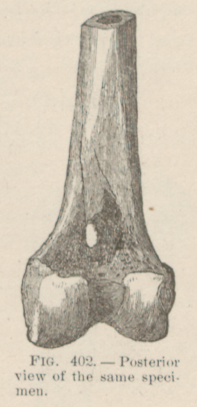 FIG. 36.—Exfoliation from the right parietal,
from gunshot contusion. Spec. 613, Sect. I, A. M.
M.
FIG. 36.—Exfoliation from the right parietal,
from gunshot contusion. Spec. 613, Sect. I, A. M.
M.