Title: Stearnes, George W.
Source text: The Medical and Surgical History of the War of the Rebellion. (1861-65.), Part 2, Volume 1 (Washington, DC: Government Printing Office, 1879), 300-301.
Civil War Washington ID: med.d1e41875
TEI/XML: med.d1e41875.xml
A case of diarrhœa of nearly two months duration:
CASE 880.—Corporal George W. Stearnes, company I, 9th New Hampshire volunteers, was mustered into service August 15, 1862, to serve three years. There is no record of the time at which he was first taken sick; but he appears on the register of the general field hospital of the 9th Corps, near Harper's Ferry, Virginia, as under treatment for diarrhœa November 5th; sent to Alexandria November 12th. He is borne on the register of the convalescent hospital at Fort Ellsworth, near Alexandria, Virginia, admitted November 16th, general debility; no disposition recorded. The register of Fairfax Seminary Hospital, near Alexandria, Virginia, shows that he was admitted to that hospital December 8th—chronic diarrhœa—transferred to Philadelphia, December 15th. He is borne on the register of the Satterlee hospital, West Philadelphia, admitted December 16th—chronic diarrhœa. December 23d, papers were made out for his discharge from service by reason of "great debility and emaciation, the result of chronic diarrhœa, which is still unchecked." Pending the approval of these papers by the proper authority, he died, December 24th, at 5 A. M. From the circumstance that his discharge papers were made out the day before his death it is to be presumed the fatal issue was unexpected. The autopsy was made during the afternoon of the same day by Professor Joseph Leidy, of the University of Pennsylvania, who forwarded the following statement to the Surgeon General's Office with the specimens: Autopsy: Age about 30. The body was rather emaciated. The chest and abdomen presented a number of faint spots of purpura. The brain was healthy. There were no pleuritic attachments, nor was there any pleurisy on either side. Lobular pneumonia was observed in the lower lobes of both lungs; the inflamed lobules were numerous—varied from the size of a marble to that of a walnut—and were in the stage of gray hepatization. Evidences of bronchitis were also observed. The heart was healthy. The stomach was exceedingly contracted. The liver was apparently sound; the gall-bladder enormously distended with green bile; the spleen small but healthy; the pancreas and kidneys sound. The intestines were inflamed throughout; in the small intestine the inflammation increased in intensity toward the ileo-cæcal valve; the agminated glands were slightly thickened and dark-red with inflammation; the solitary glands were enlarged, and looked like yellow mustard seeds sprinkled on a red ground. The large intestine was streaked and spotted with ash-color and dark-red on a nearly uniform red ground; there were also spots of ecchymosis, and the epithelium was softened.
Nos. 93 to 98, Medical Section, Army Medical Museum, are from this case. The specimens are six successive portions of the ileum. In Nos. 93 and 94 there are well-marked valvuli conniventes, and some hypertrophy of the villi, giving a velvety appearance to the surface. The solitary follicles are enlarged to the size of small pin-heads and project above the surface of the mucous membrane. In No. 94 there is a small Peyer's patch, which is a little more distinct than normal. In Nos. 95 to 98 there are no valvuli conniventes, no hypertrophy of the villi is observable, and the intestinal walls are thinner than normal. Each of these pieces presents a number of enlarged solitary follicles, which are most conspicuous in No. 98. In both 97 and 98 there is a slightly-thickened Peyer's patch. No portion of the large intestine was preserved.
The plate facing this page is a reproduction of a photograph of No. 98, which represents it of the natural size. This piece was taken from near the ileo-cæcal valve. Just above its middle is a thickened Peyer's patch, which appears as a dark elliptical spot two inches long by an inch in transverse diameter near its middle. This is crossed obliquely from above downwards by a wrinkle in the mucous membrane, produced accidentally by the method of preparing the specimen. The surface of the patch is not materially elevated above the surface of the surrounding mucous membrane, yet it is thicker than normal, and, when examined by transmitted light, appears more opaque than any of the other patches observed in this subject. The enlarged solitary follicles are scattered over the rest of the mucous surface as little pimple-like projecting tumors, which are largest and most numerous in the lower half of the piece; the largest of them, however, do not exceed pin-heads in size. Owing to the tenuity of the mucous membrane, the course of the circular fibres of the muscular coat is plainly indicated by numerous transverse lines. For the same reason the arborescent branching of the blood-vessels is also indicated in several places.
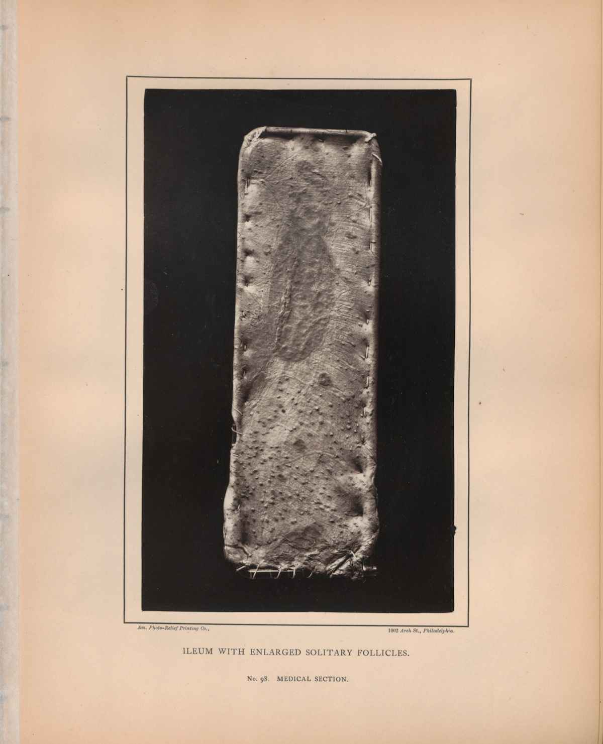 PLATE IV. ILEUM WITH ENLARGED SOLITARY FOLLICLES. No. 98.
MEDICAL SECTION.
PLATE IV. ILEUM WITH ENLARGED SOLITARY FOLLICLES. No. 98.
MEDICAL SECTION.