Title: S——, Jesse
Source text: Surgeon General Joseph K. Barnes, United States Army, The Medical and Surgical History of the War of the Rebellion. (1861–65.), Part 1, Volume 2 (Washington, D.C.: Government Printing Office, 1870), 628.
Civil War Washington ID: med.d1e20368
TEI/XML: med.d1e20368.xml
CASE.—Corporal Jesse S——, Co. B, 4th Ohio Volunteers, 3d division, Second Corps, was admitted to the McVeigh Branch of the 3d division general hospital, Alexandria, Virginia, on December 5th, 1863, with a gunshot wound of the knee-joint, received near Mine Run, Virginia, November 27th. A conical ball struck the knee of the right leg just below the patella, on its inner and anterior aspect, fracturing the inner condyle and opening the joint, passed downward and forward, and was extracted about four inches from the point of entrance. At the time of the patient's admission to the Alexandria hospital, the joint and thigh were very much inflamed and discharged pus copiously, and the man was much prostrated and terribly anxious as to the result of his wound; but after being washed and fed, and otherwise cared for, he appeared much recruited. On December 8th, the operation of excision of the knee-joint was performed by Surgeon Edwin Bentley, U. S. V.; one inch of the femur, and about one inch arid a half of the tibia bones being removed. The hæmorrhage was slight, but the condition of the tissues was not very satisfactory. The patient endured the operation well, and the reaction was good. On the 9th, the patient complained of severe pain on the right side of the chest over the lung, which continued for several days; no pain else where. On the night of December 10th, he had a severe chill, which was followed by two still more severe attacks on the 11th. Stimulants were freely given, with raw eggs well beaten up and beef essence for diet. He continued to have chills till the night of the 13th, when he was seized with a very severe chill, from which he never rallied, and died on the morning of December 14th, 1863. The post-mortem examination, eighteen hours after death, showed no attempt at reparation, and there was considerable sloughing, while upon the inside of the thigh there were numbers of small abscesses. The heart and left lung were healthy, but the right lung was infiltrated with pus, and a single large abscess appeared upon the surface of the inferior lobe. There was no pus nor abscesses found in the liver, but it was very much softened. The stomach, kidney, and intestines were perfectly healthy. The preparations in the case were sent to the Curator of the Army Medical Museum, Surgeon J. H. Brinton, U. S. V., who made the following memorandum of the appearances in the limb: "Whole joint is one suppurating mass; pus extending up and down, far down under gastrocnemius, and up between the muscles of the thigh on the inside; a long abscess in the sheath of the vessels; extending up to the middle of the thigh. Small clot in femoral artery, three inches long and thin; none in femoral vein. Surrounding tissues of vessels in some places hardened, where pus had not reached. Lining membrane of femoral vein dirty gray and softened." Clot in suphena vein. Dr. Brinton added the following notes of the preparation of the lung, represented in PLATE XIII, received at the Museum December 15th, 1863: "Apparently a metastatic abscess very circumscribed. When examined, found to be a clot, in different degrees of softening, and red blood corpuscles in every state of change; but no pus. The greenish mass in each [condyle?] was complete circumscribed gangrene of the cancellated bony tissue (coinciding with Virchow's doctrine). See picture painted by M. STAUCH." The picture is very accurately copied in the chromo-lithograph opposite,¹ PLATE XIII.
¹The preparation of the lung was preserved in the Army Medical Museum and numbered 1910 of the surgical series, but became so much decomposed after exposure by the draughtsman, that it was discarded, and the number 1910 was assigned to another specimen. There is a partly finished drawing of the femoral vessels by M. Stauch, which has been copied and elaborated by Mr. Fabre, with a view to its reproduction in chromo-lithography. The excised fragments of the knee-joint were not preserved. The adjacent portions of the diaphyses of the femur and tibia are numbered 1909, Sect, I, A. M. M., and are figured in the Catalogue, p. 336, FIG. 115. See, for a note of the case, the surgical report in Circular No. 6, S. G. O., 1865, p. 59.
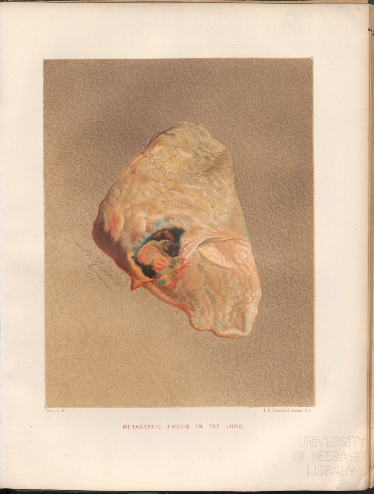 PLATE XIII. — METASTATIC FOCUS
IN THE LUNG.
PLATE XIII. — METASTATIC FOCUS
IN THE LUNG.