Title: A——, Asahel
Source text: Surgeon General Joseph K. Barnes, United States Army, The Medical and Surgical History of the War of the Rebellion. (1861–65.), Part 1, Volume 2 (Washington, D.C.: Government Printing Office, 1870), 554.
Civil War Washington ID: med.d1e19968
TEI/XML: med.d1e19968.xml
CASE 8.—Private Asahel A——, Co. F, 25th Massachusetts Volunteers, aged 25 years, was wounded at Cold Harbor, Virginia, June 3d, 1864; a conoidal ball entered the left axilla and lodged at the posterior border of the scapula. He was taken to the field hospital of the 2d division, Eighteenth Corps, where the ball was removed and simple dressings were applied. On June 11th, he was transferred to Lincoln Hospital, Washington. On the 15th, intermediary hæmorrhage to the amount of about twenty ounces occurred, probably from some branch of the axillary artery. The wound was dilated and a small artery, probably one cut in dilating the wound, ligated. The wound was filled with lint saturated in a solution of persulphate of iron, and a compress applied. The axillary artery could be distinctly felt with the finger, pulsating in the wound. Pulse regular, but weak; patient looked pale. Anodynes and stimulants were administered. On the next day hæmorrhage again occurred, which yielded to strong pressure on the compress. June 17th, 10 A. M.: Patient very pale, anæmic, and suffering from much pain in the arms and shoulder. The compress and plugging were removed, when the blood gushed out alarmingly. The wound was freely dilated and the axillary artery ligated. The hæmorrhage stopped and at the same time the heart ceased to beat. The necropsy showed the axillary largely opened about the middle of its course, on the side next to the track of the ball. There had evidently been sloughing through nearly the whole calibre of the artery. The adjacent wood-cut (FIG. 254) represents a preparation of the left axillary artery, with a large and deep ulceration, involving nearly half of the cylinder of the vessel, about an inch above the origin of the subscapularis. It was contributed, with a history of the case, by Acting Assistant Surgeon W. L. Herriman.
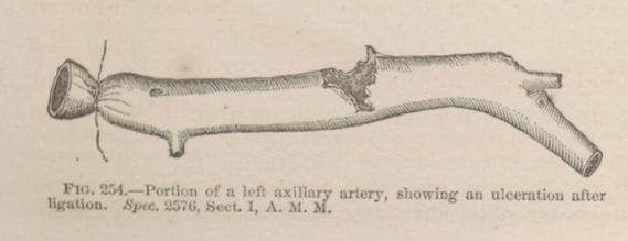 FIG. 254.—Portion of a left axillary artery, showing an
ulceration after ligation. Spec. 2576, Sect. I, A. M. M.
FIG. 254.—Portion of a left axillary artery, showing an
ulceration after ligation. Spec. 2576, Sect. I, A. M. M.