Title: M——, A——
Source text: Surgeon General Joseph K. Barnes, United States Army, The Medical and Surgical History of the War of the Rebellion. (1861–65.), Part 1, Volume 2 (Washington, D.C.: Government Printing Office, 1870), 265.
Civil War Washington ID: med.d1e16697
TEI/XML: med.d1e16697.xml
CASE.—Private A—— M——, Co. D, 155th Pennsylvania Volunteers, was wounded at the battle of Fredericksburg, Virginia, December 13th, 1862, by a conoidal musket ball, which entered at the juncture of coronal and squamous sutures, and lodged. He was admitted to the Patent Office Hospital, Washington, D. C., on the 17th, complaining but very little of his wound, and was able to walk about until the 19th, when the pain became so severe that he was compelled to take to his bed. He became restless, showing all the symptoms of febrile excitement. A lotion of lead water and laudanum was ordered to be applied to the wound, and an anodyne administered. Up to this time he had not been delirious and was able to give rational answers to all questions. On the morning of the 20th, he was comatose and all the symptoms of compression of the brain were well marked; his eyeballs, especially the right, were prominent and the pupils fixed and contracted. On removing the dressing, blood and brain substance oozed from the wound. The wound of the scalp was then enlarged and the skull trephined; spiculæ of bone were removed, causing considerable hæmorrhage, but no relief to the patient. He died a few hours after the operation. The post-mortem examination revealed cerebro-meningitis, advanced to the stage of suppuration, pus having collected over the anterior surface of the brain, and between the pia mater and arachnoid. The ball had lacerated the anterior portion of the middle lobe of the brain, the terminal branches of the internal carotid, and the anterior branch of the temporal artery. There was a large clot on the floor of the middle fossa of the cranium. A fragment of the ball was found in the centre of the middle lobe of the cerebrum, and the remaining portion was imbedded in the sphenoid bone. The pathological specimen is figured in the wood-cut. The opening in the cranial wall measures one inch from above downward and is three-fourths of an inch wide; from this point one fissure passes downward across the glenoid cavity and a second forward into the external wall of the right orbit, which is comminuted. The ball is encrusted with calcareous matter. Acting Assistant Surgeon J. H. Jamar contributed the specimen and history.
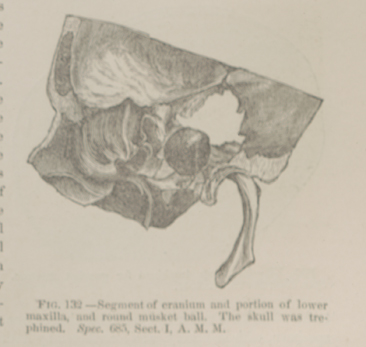 FIG. 132.—Segment of cranium and portion of lower maxilla,
and round musket ball. The skull was trephined. Spec. 685, Sect. I,
A. M. M.
FIG. 132.—Segment of cranium and portion of lower maxilla,
and round musket ball. The skull was trephined. Spec. 685, Sect. I,
A. M. M.