Title: K——, B——
Source text: Surgeon General Joseph K. Barnes, United States Army, The Medical and Surgical History of the War of the Rebellion. (1861–65.), Part 1, Volume 2 (Washington, D.C.: Government Printing Office, 1870), 264.
Civil War Washington ID: med.d1e16676
TEI/XML: med.d1e16676.xml
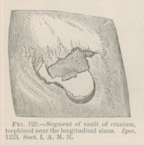
CASE.—Private B—— K——, Co. G, 6th Ohio Cavalry, aged 21 years, was wounded in a cavalry skirmish near Middleburg, Virginia, June 21st, 1863, by a carbine ball, which fractured the right parietal bone near the junction of the coronal and sagittal sutures. The bone was depressed one-sixth of an inch. A portion of the ball and several spiculæ of bone were removed on the field. The patient was conveyed to Washington, D. C., and admitted into Stanton Hospital on the 24th, being perfectly conscious, but complaining of headache. The pupils were normal, deglutition good, pulse accelerated and rather feeble, and the left lower extremity paralyzed. An ice bag was applied to the head, an enema administered, and quiet enjoined. He passed a very restless night and on the following morning became delirious, with a pulse at 120. On the 26th, coma was profound, respiration sterterous, and slower than natural, the skin hot and dry, and the pulse ranging from 65 to 80. The pupil of left eye was dilated and not responsive to the stimulus of light, that of the right eye was closely contracted, and the conjunctiva injected with blood; the left leg and arm were paralyzed, the micturition involuntary. Surgeon John A. Lidell, U. S. V., made an incision two inches in length, applied the trephine on the right edge of the fracture and cut out a disc of bone, and removed, with an elevator, two fragments of depressed bone; one, about one and a half inches in length by three-fourths of an inch in breadth, embracing both tables of the skull, the other being a small fragment of the inner table. The dura mater at the posterior and external part of the opening was found to be lacerated to the extent of half an inch, and a small quantity of brain tissue escaped. The longitudinal sinus having been uncovered, a copious stream of dark-colored blood came away, apparently flowing from the open mouths of the small veins which run from the cranium into the sinus. The bleeding was checked by a pledget of lint, saturated with a solution of persulphate of iron. The pupil of the right eye expanded to the natural size and that of the left diminished and responded to the light. The engorgement of the conjunctiva of the right eye perceptibly decreased, the stertor disappeared and the breathing became more natural; the pulse rose to 110, but consciousness did not return. Ice was again applied to the head and an enema was ordered. The next morning respiration was 60 per minute, and bronchial rattles were audible throughout the chest; pulse 130, and weak; the left side of the body was rigid, while the right side was moved quite freely. There were convulsive twitchings of right side of face, which, in two hours, extended over the entire right side of the body, while the left side lost its rigidity, but was not affected by convulsive movements. In the meantime the breathing became more frequent and feeble, and the patient died at five o'clock P. M., June 27th, 1863. At the autopsy, an elongated opening in the calvaria was exposed, half an inch long and three-fourths of an inch in width, commencing one-fourth of an inch behind the coronal suture and extending backward and a little to the right of the median line. The dura mater was lacerated to the extent of half an inch at the posterior end of the chasm in the skull. On raising that portion of the dura mater which covers the convex surface of the right hemisphere of the cerebrum, a quantity of coagulated blood was found in the cavity of the arachnoid, spread out over the convexity of the right hemisphere; the largest quantity of effused blood was found at the base of the middle and posterior lobes of the right hemisphere. The effused blood, which was very dark, amounted, in all, to three ounces, and came from the longitudinal sinus. The whole brain showed very great venous congestion. There was softening of the brain tissue at the seat of injury, near the summit of each cerebral hemisphere, but it was more marked on the right than on the left side. The pathological specimen is No. 1333, and was contributed, with the history, by Surgeon John A. Lidell, U. S. V. This case is erroneously reported as a sabre cut, in bound MSS. Div. Surg. Rec. S. G. O. No. 63, p. 22.
