Title: H——, J. C.
Source text: Surgeon General Joseph K. Barnes, United States Army, The Medical and Surgical History of the War of the Rebellion. (1861–65.), Part 1, Volume 2 (Washington, D.C.: Government Printing Office, 1870), 264.
Civil War Washington ID: med.d1e16670
TEI/XML: med.d1e16670.xml
CASE.—Corporal J. C. H——, Co. E, 2d New Jersey Volunteers, aged 37 years, was wounded at the battle of Spottsylvania Court-house, Virginia, May 14th, 1864, by a conoidal ball, which entered the lower part of the occipital bone, fractured and depressed the inner table, and lodged in the diploë, plugging up the lateral sinus on the right side of the brain. He was admitted on the same day to the hospital of the 1st division, Sixth Corps, and thence, on the 19th, sent to the Harewood Hospital, Washington, D. C. On the 21st, the parts were in a healthy condition, with a moderate amount of suppuration, and the patient's constitutional condition was remarkably good. Surgeon R. B. Bontecou, U. S. V., applied the trephine, but did not remove the ball, for fear of hæmorrhage from the lateral sinus and immediate death. The patient did not exhibit any symptoms of compression until the 26th, when he was seized with convulsions, caused by the depressed portion of bone and the pressure of the ball on the brain. The trephine was again applied, and the depressed bone was removed, but the ball had receded from sight, and could not be found. After the operation, the convulsions ceased, a passive delirium supervened and continued until the 4th of June, 1864, when death occurred. The autopsy revealed a disorganized pulpy condition of the posterior lobe of the brain, emitting an extremely offensive odor. The fracture extended from orifice of entrance to the foramen magnum. The ball was found in the posterior lobe of the cerebrum, at the depth of about two inches. The specimen is a large section of the cranium with a conoidal ball suspended in a perforation of the occipital bone. The opening measures one inch by one and one-fourth inches, and is partly caused by the operation of trephining. A fissure passes down ward and inward to the foramen magnum. The specimen and history were contributed by Surgeon R. B. Bontecou ,U. S. V. and are further illustrated in the Surgical Photograph Series, A. M. M., Volume VII, page 1.
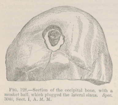 FIG. 128.—Section of the occipital bone, with a
musket ball, which plugged the lateral sinus. Spec. 3040, Sect. I,
A. M. M.
FIG. 128.—Section of the occipital bone, with a
musket ball, which plugged the lateral sinus. Spec. 3040, Sect. I,
A. M. M.