Title: H——, W——
Source text: Surgeon General Joseph K. Barnes, United States Army, The Medical and Surgical History of the War of the Rebellion. (1861–65.), Part 1, Volume 2 (Washington, D.C.: Government Printing Office, 1870), 263-264.
Civil War Washington ID: med.d1e16662
TEI/XML: med.d1e16662.xml
CASE.—Private W—— H——, Co. B, 4th Michigan Volunteers, was wounded at the battle of Fredericksburg, Virginia, December 13th, 1862, by a conoidal ball, which caused a gaping ragged wound an inch and a half long, antero-posteriorly over the left parietal, fracturing the bone. A probe could be passed through the opening so as to touch the dura mater. He was admitted to the hospital of the 1st division, Fifth Corps, and on December 16th, sent to Eckington Hospital, Washington, D. C. He was rational, but his mind was confused; the pulse was 80 and normal; the skin moderately warm; pupils somewhat contracted and fixed; some dysphagia, but no paralysis existed, and he complained of a constant tingling in his right arm and hand. On the 19th, his memory was entirely gone. The operation of trephining was decided upon; the patient was etherized, and Acting Assistant Surgeon Henry W. Fisher elongated the original wound and made another section, forming a ⊤ shaped incision. Upon turning back the flaps an extensive irregular fracture was discovered, also a small piece of bone was found driven down half its thickness below the surface. At its posterior edge a small fragment of lead was impacted. The trephine was applied and a button removed, revealing extensive comminution of the internal table toward the vertex. A tongue of bone, extending from the opening made by the trephine to a radiating fracture was removed by a Hays's saw, and two irregular fragments of the inner table, besides numerous small spiculæ, were removed. The dura mater was discolored but not lacerated save by a small puncture made by one of the spiculæ. All extraneous substances having been removed, the wound was closed and cold water dressings were applied. On December 20th, the patient was semi-comatose, but quite rational when spoken to. The next morning the pupils became contracted, the coma deepened, and all the symptoms of compression of the brain appeared. Thinking that there might be a clot under the dura mater. Dr. Fisher made a small crucial incision in the membrane, but no clot was found. The wound of the scalp and the dura mater were covered with an ash-colored, semi-fluid, sloughy matter; but on cleaning the dura mater it was found not to be sloughing, but roughened and livid. No improvement took place and the patient gradually sank until three o'clock P. M., December 21st, when he died. On removing the calvarium, the membranes were found congested, but without change of texture, save the roughening and discoloration before noted immediately about the wound. On removing the membrane, the surface of the cerebrum was found to be in a disorganized pulpy condition for a space of an inch and a half. The convolutions were obliterated, the white and gray portions being undistinguishable, and the tissue a disorganized sanious mass, so thin that several drops ran, by their own gravity, out upon the table. The rest of the brain was healthy. This disorganization was found to extend down to a level with the lateral ventricle and inward almost to the outer margin of the ventricle. The adjacent wood-cut represents the specimen, and shows the vault of the cranium, with a disk and twelve fragments removed by the trephine from the left parietal bone. The opening of the operation measures three-fourths by one and a quarter inches, and a fissure traverses the bone diagonally from the anterior superior to the posterior inferior angle. The specimen and history were contributed by Acting Assistant Surgeon S. A. Starrow.
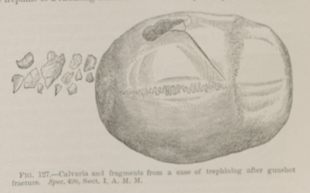 FIG. 127.—Calvaria and fragments from a case of trephining after gunshot fracture. Spec. 496, Sect. I, A. M. M.