Title: B——, W. M.
Source text: Surgeon General Joseph K. Barnes, United States Army, The Medical and Surgical History of the War of the Rebellion. (1861–65.), Part 1, Volume 2 (Washington, D.C.: Government Printing Office, 1870), 262.
Civil War Washington ID: med.d1e16641
TEI/XML: med.d1e16641.xml
CASE.—Private W. M. B——, Co. E, 83d Pennsylvania Volunteers, aged 20 years, was wounded at Petersburg, Virginia, June 20th, 1864, by a conical ball, which fractured and depressed both tables of the frontal bone just below the fronto-parietal suture and to the left of the median line. He was immediately admitted to the 1st division, Fifth Corps, hospital; on June 24th, sent to the Mount Pleasant Hospital, Washington, and thence, on the 27th, transferred to the Satterlee Hospital, Philadelphia. His pulse became slow and feeble, and on July 14th, his condition was very low. A disk of bone, one and a half inches in diameter, was removed, exposing the meninges of the brain. Pus flowed freely from the opening. The wound subsequently became gangrenous, and death ensued on July 26th, 1864. The post-mortem examination revealed an abscess in the left anterior lobe of the brain, with pus in the ventricles. The pathological specimen is figured in the wood-cut. The opening in the frontal bone is surrounded by a narrow ring of porous and diseased bone. The fractured portion of vitreous table measures one and a half by two inches. Two fragments remain in situ depressed one line. These fragments are covered by a thin, granular, mortar-like layer of calcareous matter. The specimen was contributed by Acting Assistant Surgeon G. P. Sargent.
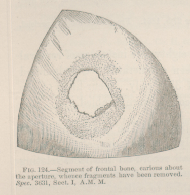 FIG. 124.—Segment of frontal bone, carious about the
aperture, whence fragments have been removed. Spec. 3631, Sect. I,
A.M. M.
FIG. 124.—Segment of frontal bone, carious about the
aperture, whence fragments have been removed. Spec. 3631, Sect. I,
A.M. M.