Title: B——, Joseph
Source text: Surgeon General Joseph K. Barnes, United States Army, The Medical and Surgical History of the War of the Rebellion. (1861–65.), Part 1, Volume 2 (Washington, D.C.: Government Printing Office, 1870), 214.
Civil War Washington ID: med.d1e14769
TEI/XML: med.d1e14769.xml
CASE.—Private Joseph B——, Co. D, 17th Massachusetts Volunteers, was wounded near New Berne, North Carolina, September 1st, 1862, by a fragment of shell which entered about the centre of the frontal bone, passed around the left side of the head, and emerged near the temporo-frontal suture. He was admitted to the Douglas Hospital, Washington, on September 5th, 1862, being partially conscious, with the right eye closed and greatly swollen. The skull between the two apertures felt soft. On September 10th, an abscess over the right eye opened about the middle of the upper lid, and pus and a few fragments of bone were freely discharged. On September 13th, the patient became comatose, and died on September 16th, 1862. At the autopsy, a large abscess was found in the anterior lobe of the left hemisphere. The pathological specimens are Nos. 236 and 514. The former shows a section of the cranium with an extensive comminuted fracture of the frontal bone a little to the left of the median line; the latter a wet preparation of the encephalon, with perforation of the dura mater, and abscess in the upper part of the anterior lobe of the left half of the cerebrum. The specimens were contributed by Assistant Surgeons W. Webster and J. W. Williams, U. S. A., respectively. The calvaria is represented in the wood-cut, (FIG. 111.) It is very thin.
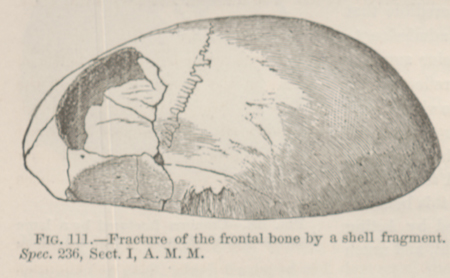 FIG. 111.—Fracture of the frontal bone by a
shell fragment. Spec. 236, Sect. I, A. M. M.
FIG. 111.—Fracture of the frontal bone by a
shell fragment. Spec. 236, Sect. I, A. M. M.