Title: W——, C. C.
Source text: Surgeon General Joseph K. Barnes, United States Army, The Medical and Surgical History of the War of the Rebellion. (1861–65.), Part 1, Volume 2 (Washington, D.C.: Government Printing Office, 1870), 181.
Civil War Washington ID: med.d1e12940
TEI/XML: med.d1e12940.xml
CASE.—Private C. C. W——, Co. I, 6th Wisconsin Volunteers, aged twenty-one years, was wounded at Spottsylvania, May 12th, 1864, and was taken to the field hospital of the 4th division of the Fifth Corps, and after the application of cold lotions to the scalp, was sent to City Point, and thence to Washington, where he was admitted into Douglas Hospital four days after the reception of the injury. Here it was ascertained that the cranium was fractured; but the symptoms were not urgent, being limited to slight paralysis of the right upper extremity, and operative interference was deferred. On May 31st a conoidal musket ball and several fragments of the left parietal were removed by Assistant Surgeon W. F. Norris, U. S. A. One large fragment of the vitreous plate was pressing on the dura mater; and this was elevated and removed. The next day, symptoms of compression of the brain were manifested. An exploration of the wound was made, and a quantity of pus was evacuated. On June 4th, 1864, twenty-three days after the injury, the case terminated fatally. At the autopsy the arachnoid was found little altered. There was an abscess in the posterior lobe of the left hemisphere, near the longitudinal sinus, of the size of a walnut, with walls of a greenish-yellow color, and communicating with the lateral ventricle. The right ventricle was filled with sero-sanguinolent fluid. There was a deposition of lymph at the base of the brain, extending from the medulla oblongata to the optic commissure. The specimen and facts connected with it were contributed by Assistant Surgeon William Thomson, U. S. A. The inner surface of the left parietal, near the fracture, is carious. Externally both parietals present over their entire surface the traces of the results of periostitis.
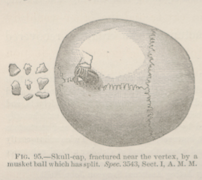 FIG. 95.—Skull-cap, fractured near the vertex, by a
musket ball which has split. Spec. 3543, Sect. I, A. M. M.
FIG. 95.—Skull-cap, fractured near the vertex, by a
musket ball which has split. Spec. 3543, Sect. I, A. M. M.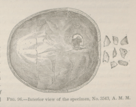 FIG. 96.—Interior view of the specimen, No. 3543,
A. M. M.
FIG. 96.—Interior view of the specimen, No. 3543,
A. M. M.