Title: Sullivan, Denis
Source text: Surgeon General Joseph K. Barnes, United States Army, The Medical and Surgical History of the War of the Rebellion. (1861–65.), Part 1, Volume 2 (Washington, D.C.: Government Printing Office, 1870), 148-149.
Civil War Washington ID: med.d1e10987
TEI/XML: med.d1e10987.xml
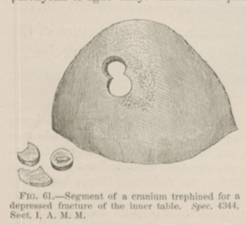
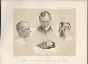
CASE 17.—Private Denis S——, Co. E, 2d Virginia Cavalry, aged 21 years, was wounded, in an engagement at Harper's Farm, near Appomattox Court House, on April 6th, 1865, by the oblique impact of a musket ball which denuded and contused the frontal bone a little below the coronal suture and to the left of the median line. Being taken prisoner, he was placed in a field hospital where a water dressing was applied, the hair being shaved off to a suitable extent. A few days subsequently, he was sent to the rear, and he reached Washington a fortnight after the reception of his wound, and was placed in Harewood Hospital on April 19th. He had a chill soon after his admission, and reported that for some days he had suffered from two paroxysms of ague daily. He had no pain in the head, nor any symptom to excite apprehension as to the condition of the brain, except the chills, which were ascribed to malarial influence. They proved, however, not to be amenable to quinia, which was freely administered, for several days, without advantage. On April 24th, a slight congestion of the lower lobe of the right lung was noticed. The next day pneumonia was fully developed here, and on the 26th the greater portion of the right lung was involved, and there was acute pain in the cardiac region, with a souffle accompanying the first sound of the heart and a murmur of regurgitation the second sound. The pulse rose rapidly to 156; but fluctuated greatly in frequency and force. At ten in the evening of this day the patient became comatose. Shortly afterwards, Surgeon R. B. Bontecou, U. S. Vols., applied the crown of a small trephine on the right of the space in which the pericranium was removed. When the outer table was passed, pus began to exude from the cells of the diploë. When this was penetrated a depressed fracture of the inner table was discovered. Another perforation was now made to obtain space to remove the depressed fragments of vitreous plate. A small fragment and another measuring nine by six lines were found completely detached, and were removed by common dissecting forceps. The operation had no influence upon the profound coma, that persisted until the patient's death, which took place on the following morning, April 28th, 1865. At the autopsy, a large abscess was found in the substance of the right cerebral hemisphere. A segment of the frontal bone, with the two disks and the larger fragment of the inner table, removed at the operation, were forwarded to the Army Medical Museum by Surgeon R. B. Bontecou, U. S. V., and are represented in the accompanying wood-cut, (FIG. 61.) A view of the interior of the specimen is given in FIG. I, of the Catalogue of the Surgical Section of the Army Medical Museum, (p. 6.) and in the Surgical Report in Circular No. 6, S. G. O., 1865, p. 11, FIG. 6, and a photograph of the patient, taken a few hours prior to the operation, is preserved at the Museum, as No. 58, Vol. I, of the Series of Contributed Photographs of Surgical Cases. The lower figure in Plate III, opposite page 105, is copied from the photograph, and represents the appearance of the patient after the graver symptoms of compression of the brain had set in.

