Title: Private Jonathan Wallace
Source text: The Medical and Surgical History of the War of the Rebellion. (1861-65.), Part 3, Volume 2 (Washington, DC: Government Printing Office, 1883), 204-205.
Civil War Washington ID: med.d2e9770
TEI/XML: med.d2e9770.xml
CASE 415.—Private Jonathan Wallace, Co. F, 21st Georgia, aged 38 years, was wounded by a conoidal ball, in the charge on Fort Steadman, March 25, 1865. The missile entered on the external surface of the left thigh at the lower portion of the middle third, passed obliquely upward and inward, fractured the femur through the middle third, and emerged on the internal surface an inch above the wound of entrance. He was taken to the field hospital of the 2d division, Ninth Corps, where Surgeon G. W. Snow, 35th Massachusetts, reports that "resection at the middle third of the femur was performed by Surgeon G. R. Sullivan, 39th New Jersey, and anterior splints applied." On April 10th, he was admitted into the Armory Square Hospital, Washington. Acting Assistant Surgeon George K. Smith reports "that the injured limb was shortened one inch; extension was applied (Buck's method) with a weight of eleven pounds, after which there was no shortening. Patient did remarkably well until June 8th, when he was attacked with diarrhœa, which was arrested at the end of three days. On June 23d, the bone appeared to be firmly united with half an inch shortening, and, on July 2d, extension was removed, and the patient rode about the ward in an invalid chair. July 28th, it was discovered that the limb had shortened two and one-fourth inches; extension was reapplied with a weight of sixteen pounds, and, on August 5th, it measured three-fourths of an inch shorter than its fellow. On August 15th, a photograph of the patient was taken [No. 92 of the Surgical Photograph Series of the Army Medical Museum, a reduced copy of which is represented in the wood-cut, FIG. 164]. On August 17th, the wound had nearly healed, his condition was remarkably good, and the bone had apparently united." On this date he was transferred to the Douglas Hospital, Washington, to the care of Assistant Surgeon W. F. Norris, U. S. A., who reports: "The wound was still open and discharging a small quantity of matter, but there was firm union of the broken femur, and the patient could without assistance raise his leg from the bed. His general health seemed to be improving, and he daily rode about the ward in a wheeled chair, the limb being supported in an extended position. August 26th several small loose fragments of bone were removed. August 28th, had a severe chill; on the 29th, had an attack of erysipelas in the thigh, and, on the following day, a loose piece of necrosed bone, one and a half inches in length, was removed. On September 2d, erysipelas had spread down to ankle joint. 11th, erysipelas had disappeared; another small sequestrum was removed from the posterior wound. 16th, had two chills, diarrhœal passages, and vomiting. 21st, he steadily grew weaker; the wound had almost ceased to discharge. There was slight icterus; the countenance was pinched and anxious and the breathing labored. 22d, he had pain in chest and abdomen, and was unable to pass his urine. These symptoms continued until the evening of the 23d, when death ensued from pyæmia. At the autopsy, fifteen hours after death, the brain appeared healthy; but there was a large amount of serous subarachnoid effusion. Both lungs were adherent and thickly studded with pyæmic patches, most of which were dark colored and hardened, a few only having softened, and containing pus. There was considerable serous effusion in the left pleural sac. The spleen was enlarged but not softened; the other thoracic and abdominal viscera appeared healthy. The fractured femur [which is represented in the adjoining wood-cut, FIG. 165] was removed and sawn longitudinally; above the fracture the marrow and interspaces between the cancelli presented a reddish chocolate hue, below, it appeared reddened and inflamed; it had not, in either locality, any gangrenous odor. The femoral vein [represented in PLATE XXX, opposite] was found to contain old and partially disintegrated blood clots, and in some portions also a quantity of healthy looking creamy pus; its walls were much thickened. It continued to present the same appearance up to about two inches below its junction with the internal iliac vein; here the clots ceased, and the coats of the vein, although of reddish hue, did not appear much thickened. The pus was carefully examined with the microscope and presented its usual round corpuscles, which under the application of acetic acid exhibited distinctly their characteristic double and triple nuclei. The femoral artery appeared to be healthy."
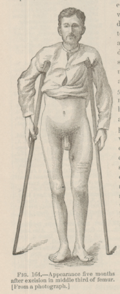 FIG. 164—Appearance five months after excision in middle third
of femur. [From a photograph.]
FIG. 164—Appearance five months after excision in middle third
of femur. [From a photograph.]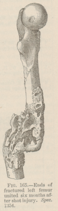 FIG. 165—Ends of fractured left femur united six months after
shot injury. Spec. 1354.
FIG. 165—Ends of fractured left femur united six months after
shot injury. Spec. 1354.PLATE XXX. — OBSTRUCTED FEMORAL VEIN