Title: Ewing, J. H.
Source text: The Medical and Surgical History of the War of the Rebellion. (1861-65.), Part 2, Volume 2 (Washington, DC: Government Printing Office, 1876), 550.
Civil War Washington ID: med.d2e31142
TEI/XML: med.d2e31142.xml
CASE 1526.—Bugler J. H. Ewing, Co. L, 8th Illinois Cavalry, aged 22 years, was wounded at Muddy Run, near Culpeper, November 8, 1863, and was taken to the Cavalry Corps Hospital, where he was operated on by Surgeon E. W. H. Beck, 3d Indiana Cavalry, who forwarded the specimen to the Army Medical Museum with the following history: "A large minié ball struck this man obliquely from the enemy's left, as he sat on his horse; his side fronting their line. The ball struck the inferior angle of the scapula, glancing upward in the direction of, and striking the neck of the humerus. About three inches of bone was broken into fragments and continuity destroyed; indeed there was the largest number of, and the smallest sized, pieces that ever came under my observation. I send you the largest pieces. It was with difficulty I got all the small fragments extricated, so deeply and firmly were they embedded in the tissues around. The ligaments were broken, and the head of the bone partially displaced from the socket; a straight incision was the only one used. No large vessels or nerves were injured by either the ball or operation. Only one small surface artery was tied; very little blood was lost. The wound was closed by sutures and straps, save the necessary aperture. Circulation was good in the arm the next morning; the patient had slept; he took nourishment, and was lively and hopeful. On the 9th, he was sent twenty-two miles in an ambulance to the Corps Hospital." The specimen consists of the head and four and a half inches of the shaft of the left humerus, excised for comminution of the upper third by a conoidal ball, which is attached, battered. The humerus was partially dislocated, but the epiphysis is uninjured. A card photograph, showing the appearance after recovery, stands with the specimen, which is represented in the accompanying wood-cut (FIG. 425). On November 10th, two days after receiving the injury, the patient was admitted to Columbia College Hospital, Washington. In the middle of January, 1864, an abscess formed in the deltoid region, and a small fragment of necrosed bone was eliminated. By the end of January, the wound was entirely healed. On March 25, 1864, the hospital report states that Ewing could slightly flex the left forearm, and that the power of pronation and supination and of moving the hand was perfect. Ewing was discharged from service September 26, 1864. On June 25, 1865, he visited theArmy Medical Museum, and a photograph was then taken, and is represented in Figure 4, PLATE XVII. He had little motion at the left shoulder joint, but the movements of the forearm were unimpaired. Examiner B. H. Long, of Mechanicsburg, Pennsylvania, November 15, 1866, reported that in consequence of the excision, "though the wound is now healed, his arm hangs helplessly at his side; no bone existing in the upper portion for a space of perhaps five inches. The arm is flexible in every direction, and requires a brace to prevent it from being a constant source of interruption." Examiner W. D. Scarff, of Bellefontaine, Ohio, September 6, 1873, states that the patient "suffers pain at times in the balance of the arm below the end of the humerus." The pensioner was paid December 4, 1873.
FIG. 425.—Excision of the upper extremity of the left humerus for shot fracture.—Spec. 1931.
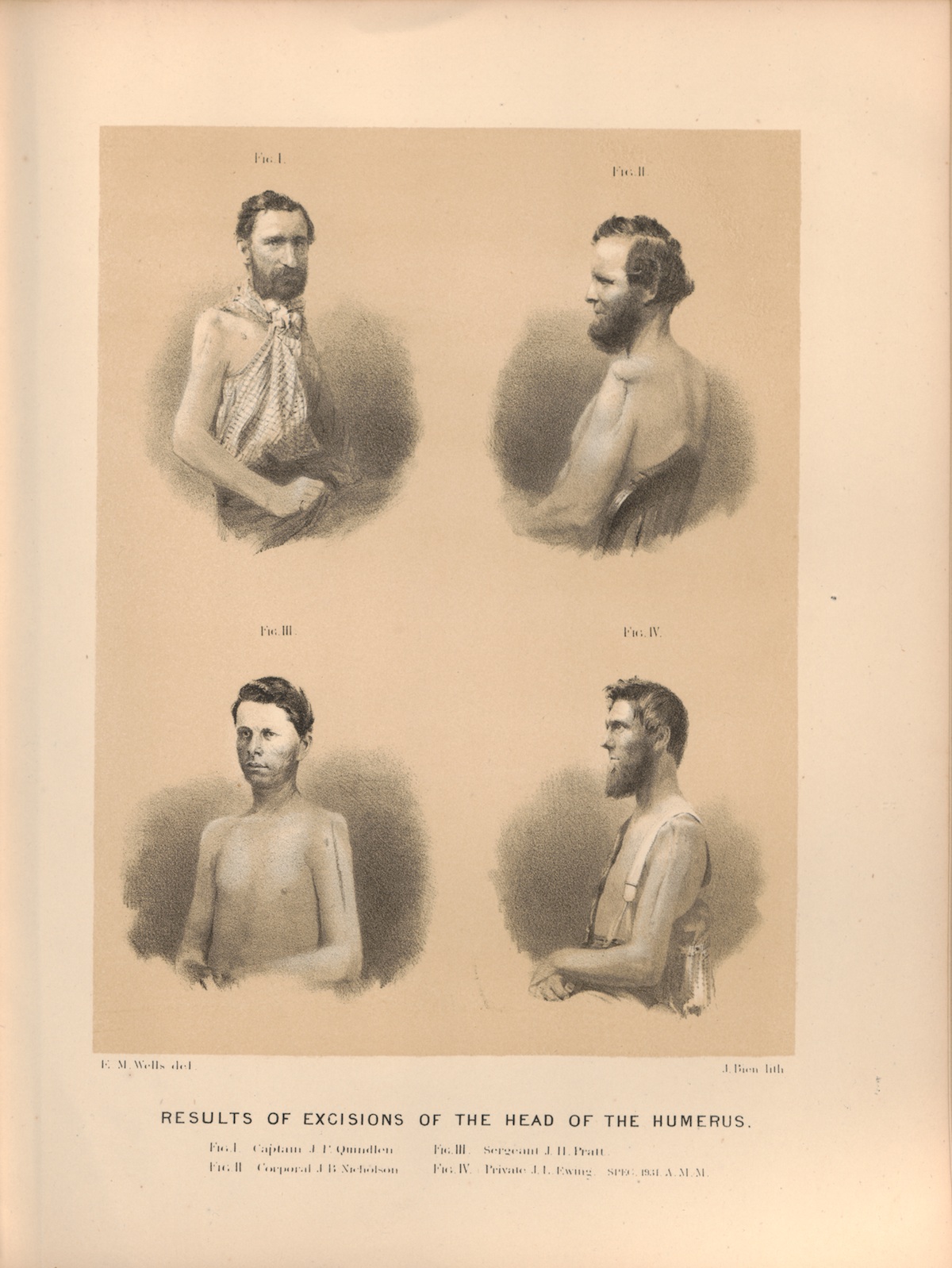 PLATE XVII. RESULTS OF EXCISIONS OF THE HEAD OF THE HUMERUS. FIG. IV. Private J. L. Ewing SPEC. 1931. A. M. M.