Title: Hay, John W.
Source text: Surgeon General Joseph K. Barnes, United States Army, The Medical and Surgical History of the War of the Rebellion. (1861–65.), Part 1, Volume 2 (Washington, D.C.: Government Printing Office, 1870), 105-106, 106.
Civil War Washington ID: med.d1e6866
TEI/XML: med.d1e6866.xml
HAY, JOHN W., Private, Co. D, 61st Pennsylvania Volunteers, aged 29 years, was wounded, at the battle of Spottsylvania, Virginia, May 11th, 1864, by a conoidal ball, which struck obliquely about the middle of the forehead. He was admitted to the hospital of the 2d division, Sixth Corps, but the injury must have been considered slight, as no record of the case was found until July 12th, when the patient was admitted to Mount Pleasant Hospital, on account of a gunshot scalp wound near the occipital protuberance, subsequently received in General Early's demonstration against the defences of Washington, the day of the patient's admission. Gangrene attacked this later wound. Bromine, nitric acid, yeast, and charcoal poultices were successively applied to the gangrenous wound. The sloughing was checked, and the wound soon assumed a healthy appearance. The wound on the forehead was not affected by gangrene, and was supposed to be trifling, and was treated with simple dressings. A month after his admission, the man complained of some pain in the forehead. Ice water was applied, and morphia was given internally. Death occurred a few hours afterwards. On August 7th, 1864, at the post mortem examination, the brain was found to be slightly congested, but no pus was observed between the skull and dura mater; yet the latter was detached from the inner table of the skull, which was carious over a surface nearly as large as the surface of the incipient exfoliation on the outer table. The scale of dead bone of the outer table remained in siter. The dura mater opposite the diseased inner table was thickened and had deposits of lymph on the surface next the cranium; otherwise, the encephalon was normal in appearance. The specimen was contributed by Assistant Surgeon C. A. McCall, U.S.A., and is represented in the foregoing wood-cuts, FIGS. 32 and 33. The notes of the case were furnished by Acting Assistant Surgeon F. J. Kern.
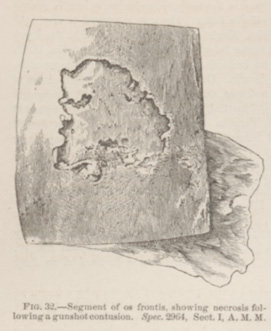 FIG. 32.—Segment of os frontis, showing necrosis following a gunshot contusion. Spec. 2964, Sect. I, A. M. M.
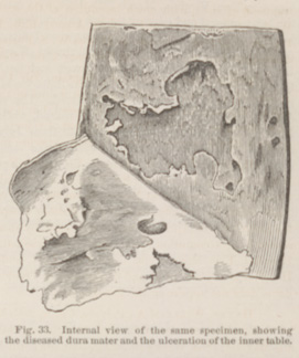 FIG. 33.—Internal view of the same specimen, showing the diseased dura mater and the ulceration of the inner table.