Title: Pierce, Henry M.
Source text: The Medical and Surgical History of the War of the Rebellion. (1861-65.), Part 2, Volume 1 (Washington, DC: Government Printing Office, 1879), 299.
Civil War Washington ID: med.d1e41868
TEI/XML: med.d1e41868.xml
The chromo-lithograph represents this very common condition, in which the pigment deposits remaining after former attacks complicated the appearances of the more recent process. The specimen was obtained from an autopsy made at the Judiciary Square hospitall, Washington, D. C., Assistant Surgeon E. J. Marsh, U. S. A., surgeon in charge, and was brought immediately to the Museum, where the water-color drawing reproduced in the plate was made by Mr. H. Faber. The following very brief history of the case was furnished:
CASE 879.—Private Henry M. Pierce, company K, 25th Maine volunteers, was taken sick while with his regiment near Chantilly, Virginia, March 23, 1863. The diagnosis on the regimental register is simply "fever." Continuing to grow worse, he was sent to Washington and was admitted to Judiciary Square hospital, April 13, 1863. Diagnosis, remittent fever. The febrile symptoms were accompanied by diarrhœa, from which he had previously suffered several times. He did well under treatment, and was apparently convalescing, though the diarrhœa still continued to the extent of several stools a day, when pneumonia supervened and was the cause of death, May 11th. Autopsy: The lower portion of both lungs was hepatized. The greater part of the ileum presented the conditions exhibited by the specimen. The mucous membrane of the colon was darkly reddened, but no marked enlargement of the solitary glands of the colon was noticed.
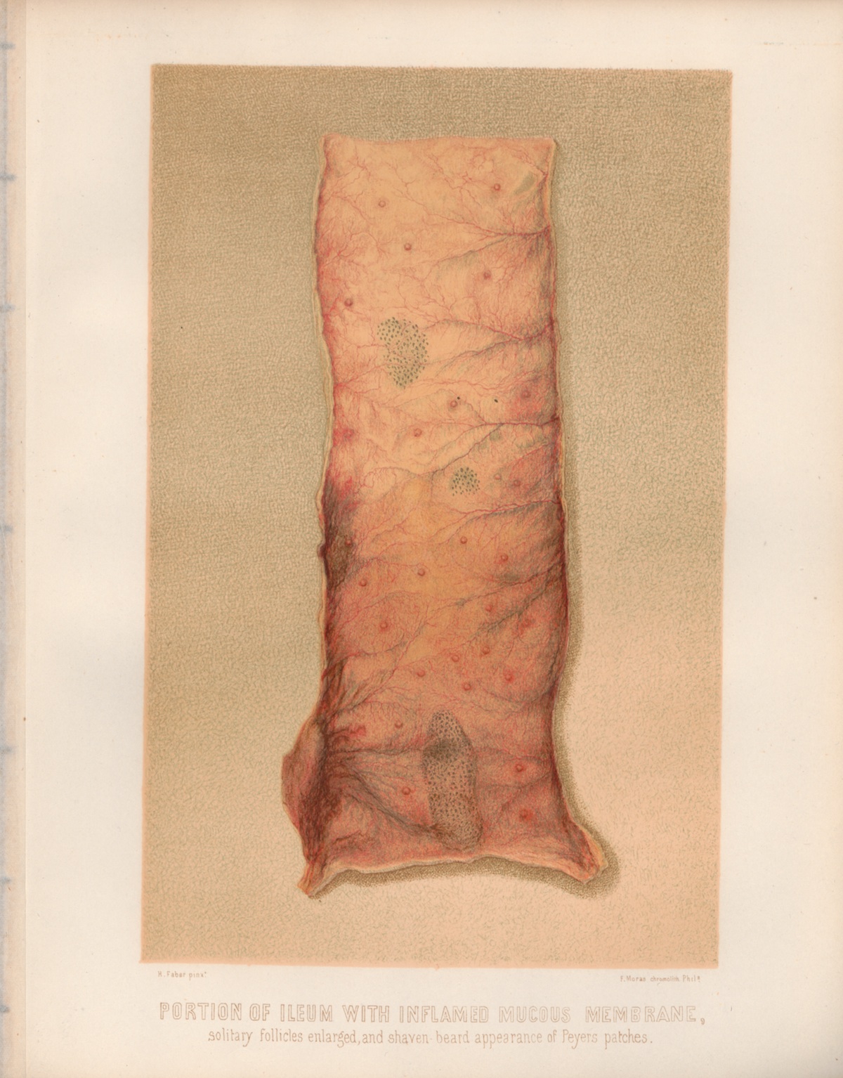 PLATE III. PORTION OF ILEUM WITH INFLAMED MUCUS MEMBRANE, solitary
follicles enlarged, and shaven beard appearance of Peyer's patches.
PLATE III. PORTION OF ILEUM WITH INFLAMED MUCUS MEMBRANE, solitary
follicles enlarged, and shaven beard appearance of Peyer's patches.