Title: R——, John
Source text: Surgeon General Joseph K. Barnes, United States Army, The Medical and Surgical History of the War of the Rebellion. (1861–65.), Part 1, Volume 2 (Washington, D.C.: Government Printing Office, 1870), 59.
Civil War Washington ID: med.d1e3617
TEI/XML: med.d1e3617.xml
CASE.—Private John R——, Co. H, 2d Michigan Volunteers, aged 41 years, was wounded on July 17th, 1865, in a street affray, receiving four wounds of the head from stones thrown at him. He was admitted to Armory Square Hospital, Washington, D. C., on the following day. He was perfectly conscious, yet had marked contraction of the pupils, with accelerated pulse, and a tremulous voice. There was considerable ecchymosis about the orbits. The first wound examined was over the frontal eminence, and penetrated no further than the aponeurosis of the occipito-frontalis muscle. The second was in the centre of the coronal suture, and slightly denuded the pericranium. The third was in the right temporal region, and likewise was a scalp wound. The fourth was on the right parietal eminence; and, upon a close examination, it was discovered that a minute depression of the bone, half an inch in diameter, existed, evidently produced by a blow from the sharp edge of the stone. The patient was a stout, muscular man, in good health; he suffered no nausea, and little pain. He was immediately placed under the influence of ether, and Surgeon D.W. Bliss, U. S. V., after shaving the scalp, made a crucial incision three inches in length, having the wound at the intersection of the incisions, and then, reflecting the flaps, applied the crown of a trephine and removed a disk of bone, which was found to include, with remarkable exactness, a depressed fragment of the vitreous plate. Between the diploe and depressed lamina there was a coagulum. The dura mater was uninjured. The wound was partly closed by four sutures, an opening being left over the perforation, into which a pledget of charpie was inserted. The patient recovered favorably from the anæsthetic, and was put to bed and ordered to observe perfect quiet and strict diet. The case proceeded without an unfavorable symptom. On July 23d, the sutures were removed. On July 24th, the compress of charpie was taken away, and a healthy granulating surface appeared beneath. These facts in regard to the case were reported by Assistant Surgeon Charles A. Leale, U. S. V. The pathological specimen was presented to the Army Medical Museum by the operator, and is represented in Photograph No. 87 of the Surgical Section of the Army Medical Museum, and in the accompanying wood-cut, (FIG. 24.) The disk is seven-eighths of an inch in diameter, and is slightly reduced in the illustration. On August 24th, 1865, the patient was transferred to Harper Hospital at Detroit, Michigan. The case continued to progress favorably, and the man recovered without a bad symptom. He was discharged from service on September 8th, 1865.
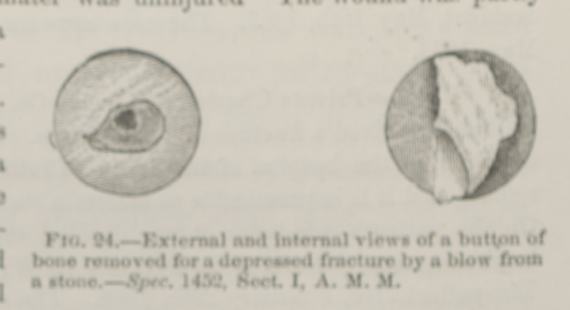 FIG. 24.—External and internal views of a button of
bone removed for a depressed fracture by a blow from a stone.—Spec. 1452, Sect. I, A. M. M.
FIG. 24.—External and internal views of a button of
bone removed for a depressed fracture by a blow from a stone.—Spec. 1452, Sect. I, A. M. M.