Title: B——, S.
Source text: Surgeon General Joseph K. Barnes, United States Army, The Medical and Surgical History of the War of the Rebellion. (1861–65.), Part 1, Volume 2 (Washington, D.C.: Government Printing Office, 1870), 490.
Civil War Washington ID: med.d1e19420
TEI/XML: med.d1e19420.xml
CASE.—Private S. B——, Co. A, 83d New York, having been wounded at Fredericksburg, December 13th, 1862, was admitted into Lincoln Hospital, Washington, on December 23d, 1862. A conoidal musket ball had entered the right chest posteriorly, over the attachment of the eleventh rib, and passed forward. The missile was extracted on the 26th, and simple dressings were applied. Pleuro-pneumonia ensued, and resulted in empyema. On January 3d, 1863, a pint of pus was evacuated from the pleural cavity. The case terminated fatally on January 21st, 1863. Nine hours subsequently a post-mortem examination was performed by Assistant Surgeon G. M. McGill, U. S. A. Rigor mortis was well marked. The brain weighed forty-five ounces and two drachms. A healthy fluid was found in the lateral ventricles. The bronchial glands were enlarged. The right lung was compressed and crowded into the superior, posterior, and internal part of its chamber. It was adherent to the costal parieties by fibrinous bands. Anteriorly, inferiorly, and externally, occupying the cavity left by the retreating lung, was found a collection of pus, between the two walls of pleura, measuring one pint and a half. This cavity was lined by a thick membrane presenting internally a mucoid appearance. The left lung showed gray hepatization in the upper and posterior portions, the inferior and anterior being congested, and thick, tenacious bronchial secretion exuded from the bronchial tubes. Anteriorly, this lung was firmly adherent to the pericardium. The right lung weighed fourteen, the left twenty-six ounces. The pericardium was filled with two ounces of yellowish fluid. The heart, with the pericardium, weighed ten and a half ounces. Firm fibrinated clots existed in both auricles, continuous within the auriculo-ventricular openings, and a black clot was found in the left ventricle and aorta. The liver, weighing sixty-four ounces, was "nutmegged." Each kidney weighed five and a half ounces. The spleen was firm, and weighed seven and a half ounces. The pancreas weighed four ounces. The greater omentum extended a short distance below the umbilicus; the intestines were much inflated; the lymphatics of the lumbar region and the mesenteric glands were enlarged. The stomach and duodenum were normal; patches of congestion were scattered through the jejunum and ileum; the mucous membrane of the small intestine was softened; the walls of the large intestine were thickened. Portions of the eleventh and twelfth ribs of the right side, completely fractured and surrounded at the points of solution with large irregular formations of callus, were contributed to the Army Medical Museum by Surgeon H. Bryant, U. S. V., and the particulars of the autopsy were furnished by Assistant Surgeon J. Cooper McKee, U. S. A. The specimens are figured in the accompanying wood-cuts (FIGS. 232 and 233), on a much reduced scale, the cut used in Circular 6, and in the Catalogue, being utilized.
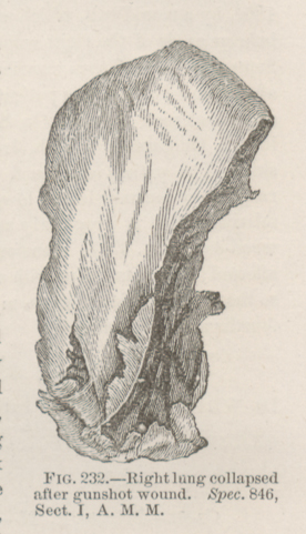 FIG. 232.—Right lung collapsed after gunshot wound. Spec. 846, Sect. I, A. M. M.
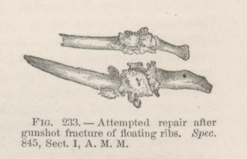 FIG. 233.—Attempted repair after gunshot fracture of
floating ribs. Spec. 845, Sect. I, A. M. M.
FIG. 233.—Attempted repair after gunshot fracture of
floating ribs. Spec. 845, Sect. I, A. M. M.