Title: H——, P.
Source text: Surgeon General Joseph K. Barnes, United States Army, The Medical and Surgical History of the War of the Rebellion. (1861–65.), Part 1, Volume 2 (Washington, D.C.: Government Printing Office, 1870), 263.
Civil War Washington ID: med.d1e16656
TEI/XML: med.d1e16656.xml
CASE.—Private P. H——, Co. E, 9th Pennsylvania Reserves, aged 28 years, was wounded at the battle of Fredericksburg, Virginia, December 13th, 1862, by a conoidal ball, which fractured and depressed the posterior portion of the left parietal bone. He was unconscious for some time after the reception of the injury. On December 15th, he was admitted into the Stanton Hospital, Washington. D. C. A wound of scalp was discovered through which could be felt a portion of depressed bone; through another wound, two inches anteriorly, the patient stated that a portion of the ball had been extracted. No symptoms of brain lesion were manifest, the patient being perfectly conscious and answering all questions correctly and intelligently. Ice was applied to the head, but, toward evening, convulsions ensued followed by others at intervals during the night. The next morning a semi-comatose condition supervened, continuing until the 17th, when it deepened. The power of deglutition was lost, and the tongue turned to the left side of the month. On the 18th, the condition being unimproved, an operation was decided upon. Accordingly the patient was etherized, and Surgeon John A. Lidell, U. S. V., applied the trephine to the anterior edge of the opening in the cranium and removed a disc of bone. The depressed portion, about an inch long and three-fourths of an inch wide, and six fragments of bone were easily detached. One of the latter had evidently perforated the dura mater, its removal being followed by a discharge of disorganized brain substance. After the operation, the coma lightened somewhat and power of deglutition and consciousness were restored; the loss of speech, however, continued. The improvement was of short duration, for on the next day coma and paralysis of the right side again supervened, with effused blood, which was very dark, amounted, in all, to three ounces, and came from the longitudinal sinus. These symptoms increased in gravity continuously, the patient becoming more and more exhausted until the 23d, when he died. At the post-mortem examination, a thick brown-colored pus, to the amount of an ounce and a halt, escaped from between the dura mater and the brain. Under the seat of injury was found red softening and disorganization of the brain extending to the depth of an inch and a half. There was also considerable effusion of clear serum in the ventricles, and a sero-sanguinolent effusion at the base of the brain. The cerebrum generally, including both right and left hemispheres, was congested, the punctiform spots being unusually distinct. The dura mater, covering the convexity of the left hemisphere, showed marks of recent inflammatory action, being injected, reddened, roughened, and of a brown color in the neighborhood of the fracture. It was also lined by a thick layer of false membrane. The specimen is figured in the wood-cut. The fractured portion of the inner table of the cranium measures three-fourths by one inch, and is partly included in the disk removed by the trephine. The outer table is injured to a less extent. The specimen and history were contributed by Surgeon J. A. Lidell, U. S. V.
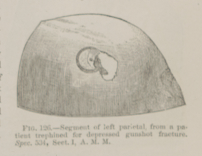 FIG. 126.—Segment of left parietal, from a patient
trephined for depressed gunshot fracture. Spec. 534, Sect. I, A. M.
M.
FIG. 126.—Segment of left parietal, from a patient
trephined for depressed gunshot fracture. Spec. 534, Sect. I, A. M.
M.