Title: D——, Ross
Source text: Surgeon General Joseph K. Barnes, United States Army, The Medical and Surgical History of the War of the Rebellion. (1861–65.), Part 1, Volume 2 (Washington, D.C.: Government Printing Office, 1870), 160.
Civil War Washington ID: med.d1e11172
TEI/XML: med.d1e11172.xml
CASE.—Sergeant Ross D——, Co. B, 19th Massachusetts Volunteers, was wounded in the engagement at Bristow Station, Virginia, October 14th, 1863, by a conoidal ball which struck near and external to the left frontal eminence, slightly depressing the external and fissuring the internal table. He was admitted to the hospital of the 2d division, Second Army Corps, and on October 19th was sent to Lincoln Hospital, Washington. No cerebral symptoms existed for some time after admission. On November 6th, hæmorrhage, which was arrested by ligation, occurred from the anterior temporal artery. Hæmorrhage recurred on November 20th, and on the following day the wound became gangrenous. The patient grew comatose and died on November 29th, 1863. At the autopsy the external table was found necrosed, the diploë was filled with fungous granulations. The dura mater was indurated beneath the injured spot, although no evidences of inflammation were present. On removing the brain a large quantity of thin pale serum was found in the subarachnoid space. A large abscess existed in the anterior of the left hemisphere just beneath the seat of injury, extending into the lateral ventricle, filled with thick, sanious and fœtid pus. The right ventricle was normal. The pathological specimen is figured in the cut (FIG. 69.) The inner table of the cranium presents a T-shaped fissure without depression, and is spongy. A thin plate of bone one inch in diameter is necrosed on the external table, and the adjacent osseous tissue is porous and cribriform. The specimen was contributed by Assistant Surgeon H. Allen, U. S. A.
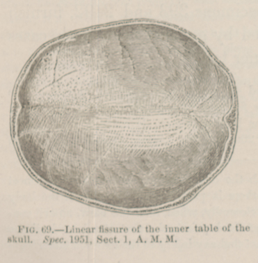 FIG. 69.—Linear fissure of the inner table of
the skull. Spec. 1951, Sect. I, A. M. M.
FIG. 69.—Linear fissure of the inner table of
the skull. Spec. 1951, Sect. I, A. M. M.