Title: Phelan, John
Source text: The Medical and Surgical History of the War of the Rebellion. (1861-65.), Part 3, Volume 2 (Washington, DC: Government Printing Office, 1883), 107-108.
Civil War Washington ID: med.d2e5443
TEI/XML: med.d2e5443.xml
CASE 250.—Captain John Phelan, Co. A, 73d New York, aged 22 years, received a compound comminuted fracture of the neck and upper extremity of the left femur at the battle of Spottsylvania Court House, on May 14, 1864. On May 16th, he was admitted to Mount Pleasant Hospital, Washington. The rapid and incessant influx of wounded was such that the attention of the overworked hospital staff was not especially drawn to his case for some time after his admission, and the delay in minutely examining the case was extended by the uncomplaining fortitude of the sufferer, who expressed his wish that the more serious cases should first be attended to, and declared that his own sufferings were comparatively slight. When, however, Acting Assistant Surgeon Mulford, the ward surgeon, proceeded, on June 3d, to adapt apparatus to what he supposed to be an ordinary gunshot fracture of the upper third of the thigh, he was led to apprehend that the injury extended to the coxo-femoral articulation, and requested the opinion of the surgeon in charge of the hospital, Assistant Surgeon C. A. McCall, U. S. A., as to the diagnosis and treatment. Dr. McCall immediately visited the patient, and found him to be a large, muscular, finely formed man, whose previous health had been excellent. When lying quietly in bed, he suffered but little. His appetite was good; and his strength, so far, had diminished but little. Altogether, his general condition was extraordinarily good, in view of the gravity of the injury he had sustained. The ball had entered in front, just over the point at which the profunda is given off from the left femoral artery. The aperture of entrance was small and characteristic as an entrance wound of a conoidal musket ball. The missile had passed toward the great trochanter and shattered it. Further, its course could not be ascertained at the time. Any movement of the limb caused extreme pain. Though the femur was much comminuted, Dr. McCall was not positive that the hip joint was implicated, and, with a view to a full exploration of the injury, he directed Dr. Mulford to make a longitudinal incision three inches in length over the trochanter, to explore the parts thoroughly, and to ascertain by digital examination the condition of the articulation. If it was uninjured, Dr. Mulford was instructed to extract detached fragments of bone and foreign matters, to close the upper part of the wound, and to avail of the lower portion for drainage. In the afternoon the patient was etherized, and the exploratory incision was made, and it was found that the fracture extended to the head of the femur. It was then decided to excise the head. The patient was again rendered insensible by the inhalation of sulphuric ether, and Dr. McCall extended Dr. Mulford's incision upward an inch or more, and then made an oblique incision across its upper extremity, as represented in the accompanying figure (FIG. 61). The two flaps thus marked out were reflected, and the joint was readily exposed, the round ligament divided, and the head of the femur exarticulated. The acetabulum was carefully examined and found to be uninjured. Seven large and numerous small fragments of the neck and trochanter major were then removed, a task requiring much time and patience, many fragments being driven into the gluteal muscles, or deeply retracted by the muscles attached to the great trochanter. The fractured upper extremity of the femur was then brought out at the wound, by adducting and pushing upward the knee of the injured limb, and all diseased tissue was removed. The periosteum was in a healthy condition quite up to the end of the bone. The wound was now thoroughly washed out, and approximated by three stitches, and by adhesive strips. A grain of sulphate of morphia was administered, and the patient was put to bed. The operation lasted three-quarters of an hour. Dr. McCall thinks that the ball was removed during the operation; but is not positive on this point. The hospital report, which is quoted at page 69 of Circular No. 6, S. G. O., 1865, states that the patient's pulse was quick and irritable at the time of the operation, that he had a furred tongue and diarrhœa, and was reduced by suppuration. But Dr. McCall (letter of February 11, 1868) thinks that this report exaggerates the gravity of the constitutional symptoms, and is quite sure that the general condition was favorable. The patient rallied well from the operation. For two days the wound was dressed with lint. Suppuration then commencing, the limb was placed in Fergusson's apparatus for excision of the head of the femur, the counter-extension straps being left off. The wound was freely syringed with cold water containing a little permanganate of potassa. A nourishing diet was ordered, with tonics and stimulants. For a week or ten days subsequently, the case progressed favorably. Suppuration was moderate in amount, and of a healthy character. About the middle of June, the weather became intensely hot. The atmosphere of the wards, in which nearly every bed was occupied by a patient with suppurating wounds, became intensely oppressive. About this time, the patient began to grow worse. The cheerful resolution and hopefulness he had hitherto evinced, gave way. Diarrhœa supervened, and he lost strength rapidly. The fatal event was thought to have been delayed by the plan which was pursued of daily removing the patient in his bed at nine in the morning to a spot beneath the shade trees near the hospital, where he had pure air and escaped the distressing scenes of the ward; he remained each day until five in the afternoon. He died on June 21, 1864. The portion of bone excised was forwarded at the time of the operation to the Army Medical Museum. The preparation is No. 2618 of the Surgical Series. It is represented in the adjacent wood-cut (FIG. 60).
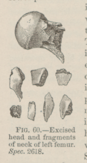 FIG. 60.—Excised head and fragments of neck of left femur. Spec. 2618.
 FIG. 61.—Direction of the incisions in case of excision of
the head of the femur. [From a drawing by Dr. McCALL.]
FIG. 61.—Direction of the incisions in case of excision of
the head of the femur. [From a drawing by Dr. McCALL.]