Title: N——, Allen A.
Source text: The Medical and Surgical History of the War of the Rebellion. (1861-65.), Part 2, Volume 2 (Washington, DC: Government Printing Office, 1876), 133.
Civil War Washington ID: med.d2e31530
TEI/XML: med.d2e31530.xml
CASE 362.—Sergeant Allen A. N——, Co. D, 2nd Massachusetts, died, in an ambulance carriage, while being conveyed to Lincoln Hospital, Washington, June 10, 1863. He had probably been wounded at Beverly Ford the day previously. An autopsy was made seven hours after the reception of the cadaver. The wound was ten inches above the pubis and three inches to the right of the median line, and was a small opening, depressed and blackened around the edges. The small intestine and omentum were agglutinated together by a thin layer of recent lymph. The cavity of the abdomen contained a large quantity of blackish fluid, mingled with dark clots of venous blood. The mucous lining of the trachea was very pale, and was covered with numerous papillated points, which were readily removed with the finger, and probably consisted of a tenacious mucous secretion. A large, dense, venous clot was present in the right ventricle of the heart, and a much smaller and more fibrous one in the left. One inch to the right of the suspensory ligament of the liver was a stellate opening, and on the inferior surface, to the left of the gall bladder, a large irregular fissure, through which the ball had passed; it went through the body, making its exit two inches to the left of the spine and six inches above the sacrum. The intestines were not injured. The specimen represented in the wood-cut (FIG. 103) was contributed to the Army Medical Museum by Surgeon G. S. Palmer, U. S. V., and the notes of the case by Assitant Surgeon H. Allen, U. S. A., who conducted the post-mortem examination.
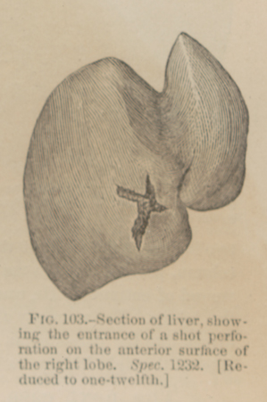 FIG. 103.—Section of liver, showing the entrance of a shot perforation on the anterior surface of the right lobe. Spec. 1232. [Reduced to one-twelfth.]