Title: Martin, M.
Source text: The Medical and Surgical History of the War of the Rebellion. (1861-65.), Part 3, Volume 2 (Washington, DC: Government Printing Office, 1883), 583.
Civil War Washington ID: med.d2e21769
TEI/XML: med.d2e21769.xml
CASE 830.—Corporal M. Martin, Co. D, 28th Massachusetts, aged 27 years, was wounded at Hatcher's Run, March 25, 1865. Surgeon F. M. Hammond, 126th New York, reported his admission to the field hospital of the 1st division, Second Corps, with "shot wound of left foot" Ten days after the reception of the injury the patient was transferred to Douglas Hospital, Washington, whence Assistant Surgeon W. F. Norris, U. S. A., contributed the pathological specimen shown in the annexed wood-cut (FIG. 334), with the following history: "The wound penetrated the ankle joint. The ball entered in front, about midway between the malleoli, and made its exit posteriorly and a little above the external malleolus, comminuting in its course the lower ends of the tibia and fibula. There was marked constitutional disturbance, headache, high fever, rapid pulse, and loss of appetite. But little swelling or inflammation in the joint, however, took place, and not much discharge. The day subsequent to the patient's admission the joint was carefully examined, when both bones were found to be much comminuted and amputation was decided as the only resort giving a fair chance of recovery. The patient, however, positively declined all operative interference. Towards evening he had a slight chill. On April 7th there was nausea and vomiting, and on the following day he had three chills, followed by fever and profuse sweats. Nausea and vomiting continued, the fluid ejected being tinged with bile; the discharge from both wounds, however, retained its healthy appearance. On April 9th, 10th, 11th, and 12th the patient had a chill each day, and on the latter day there was marked yellowness of the face and conjunctivæ; slight cough and delirium. On April 13th there was another chill; pulse 110, respiration 38; severe pleuritic pain in the right side of the chest. Death occurred on April 14, 1865. Rigor mortis was well marked at the autopsy, also the yellow hue of the skin and conjunctivæ. On opening the thoracic cavity nearly a pint of intensely yellow fluid was found in the right pleural cavity. Each lobe of the right lung presented numerous patches varying in size from half an inch to two inches in diameter, most of which on incision gave exit to pus; the left lung contained similar patches in both lobes. The spleen was much softened, being almost semifluid in consistence; liver enlarged but apparently healthy; other thoracic and abdominal viscera normal." The specimen (No. 253) consists of the astragalus and the lower halves of the tibia and fibula of the injured limb, the extremities of both bones of the leg being shattered.
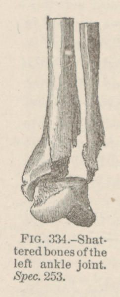 FIG. 334.—Shattered bones of the left ankle joint. Spec. 253.