Title: Gray, N.
Source text: The Medical and Surgical History of the War of the Rebellion. (1861-65.), Part 3, Volume 2 (Washington, DC: Government Printing Office, 1883), 371.
Civil War Washington ID: med.d2e15185
TEI/XML: med.d2e15185.xml
CASE 549.—Private N. Gray, Co. H, 4th Maine, aged 24 years, was wounded in the right knee joint, at the Wilderness, May 5, 1864, and entered Harewood Hospital, Washington, three weeks afterwards. Surgeon R. B. Bontecou, U. S. V., contributed the following history: "The ball entered in the middle of the internal condyle three-fourths of an inch above the inferior margin, passing in an oblique direction inward and downward. At the time of his admission the general state of his health was good and color of skin normal; knee joint a little swollen. The patient complained of a fixed pain right below the centre of the patella, which was increased by pressure upon the patella. The thigh was swollen, especially on the inner side, about five inches upward, and fluctuating. On enlarging the opening upward a quantity of pus was discharged and the ball was discovered and removed. The hole made by the ball was one inch deep; its direction inward and downward. An ice bag was applied to the joint. On June 2d, an incision was made in the fossa poplitea three inches long, and the lower third of the posterior side of the femur was found to be surrounded with pus, but the bone was yet covered with periosteum. The discharge was free and copious; the ice was still kept on. On July 20th, there was œdema of the right leg, which was bandaged from the toes to the knee joint; discharge healthy and copious. Solution of chlorinate of sodæ was now used in the dressing. By September 1st, the wound was doing well, discharging but little, and the incision in the fossa poplitea being healed. Exudation below the patella had disappeared, and the movements of the knee joint were perfectly free and painless. The patient went on furlough, and returned on September 14th. Small pieces of bone were coming from the wound in the condyle; movement of the joint perfect. On December 1st, erysipelas of the right leg commenced below the patella, attended with high fever, vomiting, and headache. For this ten drops of muriated tincture of iron were given every three hours. By December 10th, the erysipelas had disappeared and the patient was doing well. The process of exfoliation was going on slowly on January 1, 1865. On February 13, 1865, the patient was discharged from service, the process of exfoliation not yet having terminated, but the knee joint being of normal size and color, and its movements perfectly free and painless in every direction." Examiner I. H. Harding, of Ellsworth, Maine, February 1, 1867, certified to the injury and added: "The joint and leg are weakened and atrophied so much that he walks quite lame." The Boston Examining Board reported, September 13, 1875: "There are two large adherent cicatrices in the popliteal space, which impair the motions of the hamstrings." The pensioner was paid December 4, 1879. The wood-cut (FIG. 222) is a copy of a photograph contributed by Surgeon Bontecou (Card Photographs, Vol. 3, p. 22).
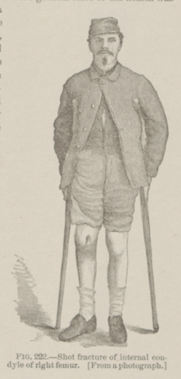 FIG. 222.—Shot fracture of internal condyle of right femur.
[From a photograph.]
FIG. 222.—Shot fracture of internal condyle of right femur.
[From a photograph.]