Title: Kelcher, Thomas
Source text: The Medical and Surgical History of the War of the Rebellion. (1861-65.), Part 2, Volume 1 (Washington, DC: Government Printing Office, 1879), 513, 520.
Civil War Washington ID: med.d1e41918
TEI/XML: med.d1e41918.xml
A very similar specimen to that just described is represented in the plate facing page 514, which is reproduced from a photograph of No. 602, Medical Section, and represents the specimen after it had been preserved in alcohol for some time. The following history of the case was furnished by Acting Assistant Surgeon George K. Smith:
CASE 900.—Private Thomas Kelcher, company E, 28th Massachusetts volunteers; age 29; Irish; enlisted August 1, 1864. March 25, 1865, he received a gunshot fracture of the left thigh in action near Petersburg, Virginia. He was admitted to the depot field hospital of the Second Army Corps, City Point, Virginia, and on the 31st was sent on board the steamer State of Maine for transfer to Washington. He was admitted to Armory Square hospital, Washington, D. C., April 1st. On examination it was found that the ball had entered the external surface of the left thigh at the junction of its middle and lower thirds, and passing transversely inwards comminuted the femur, and made its exit on the internal surface opposite the wound of entrance. April 2d, he was etherized, the wound of entrance enlarged, and five fragments of bone removed. When admitted to Armory Square this man had an abscess at the lower border of the left popliteal space, caused by the pressure of the end of a splint which had been applied to the posterior surface of the thigh. This caused so much pain and soreness that he could not at first bear extension, but at the end of the month of April this trouble was so much better that he was able to bear extension well. May 10th, he was attacked with the stools containing mucus but no blood, and this continued until his death, with the exception of two or three short intervals. June 15th, he was doing remarkably well; appetite good, tongue clean and bowels regular; but, July 1st, he had a return of the dysentery, which was not subsequently benefited by treatment. July 25th, the injured limb was attacked by erysipelas, and for a little distance around the wound, not exceeding half an inch at any point, it presented the appearance of gangrene. This was treated by the application of solution of bromine. The limb was considerably swollen, and the patient felt great pain near the ankle joint. July 29th, a large patch of the epidermis over the left ankle was raised, and the space beneath filled with dark-colored serum. The true skin presented the bluish color seen in the first stage of gangrene. This appearance continued to extend up the limb until his death, on the following day. The night before his death he had a severe pain in his left side, indicative of pleurisy. Autopsy 8 hours after death: Rigor mortis well marked. The apex of each lung was studded with tubercles. The posterior surface of the lower part of the left lung was covered with lymph, and the pleura at this point was highly inflamed. The heart was normal; the stomach and small intestine were also normal. The mucous membrane of the large intestine throughout its whole extent presented numerous ulcers about a quarter of an inch in diameter, and in the descending portion there were a large number of cysts containing lymph, some of which appeared to be undergoing ulceration, while others, already perforated, were filled with pus. The femoral and iliac veins of the injured side were completely filled with a firm fibrinous clot extending from the knee to the vena cava ascendens. The fractured femur was firmly united, with three-quarters of an inch shortening. [Specimens Nos. 602 and 603, Medical Section, are portions of the colon in this case, and No. 1105, Surgical Section, is the fractured femur.]
The large intestine from this case was brought to the Museum immediately after the post mortem examination. When received its mucous surface was of a pale cream color, with livid bluish discoloration around the cysts. These contained a semi-transparent yellowish matter resembling calves'-foot jelly, in which, with the microscope, a few delicate nucleated cells resembling the white corpuscles of the blood were observed to be imbedded. This substance was undoubtedly what is described as pus in the history of the case given below. Many of the cysts had ruptured and their contents escaped, leaving cavities resembling follicular ulcers. I found no other ulcers in any part of the specimen. The specimen, No. 602, Med. Section, represented in the photo-plate, is a portion of the transverse colon, exhibiting some fifty of the cysts described; the action of the alcohol has caused them to collapse, but their position and dimensions can still be recognized; several have so ruptured as to be converted into the cavities just described. No. 603, Medical Section, is a portion of the descending colon from the same case; it contains a small number of similar cysts, and also presents a number of small, irregular, oval cicatrices, such as result from the healing of follicular ulcers. A few of these cicatrices can also be recognized in the photo-plate by the help of a lens.
In both the cases just described the death of the patient was undoubtedly hastened by the gunshot wound.
The plate facing page 528 gives an excellent idea of the appearances presented by these characteristic cicatrices. It is reproduced from a photograph of a portion of No. 603, Medical Section. The specimen represented is from the case of Thomas Kelcher,. related on page 513. In that case the patient, while under treatment for a gunshot fracture of the thigh, suffered an attack of acute catarrhal dysentery which passed into a chronic flux. His colon presented a number of cysts, containing a jelly-like substance; these were fully described in the place referred to, and are represented in the plate facing page 514, which is reproduced from a photograph of No. 602, Medical Section. But between these cysts and the ulcer-like cavities formed here and there by their rupture there were a considerable number of stellate cicatrices. Several of these cicatrices can be recognized in the plate just mentioned as representing No. 602, Medical Section. The photograph of a portion of No. 603, reproduced in the plate now described, was taken one-half larger than nature in order that the surface details might be better seen. In the portion reproduced there are several small, deep, oval cavities which represent the ruptured cysts just referred to. The surface of the intestine exhibits, besides, rather more than a dozen stellate cicatrices, some four or five of which form a conspicuous group near the centre of the plate. Each cicatrix consists of a central smooth area, oval or almost linear in shape, and varying in the specimen from one-twentieth to one-fifth of an inch in long diameter. From this central smooth portion radiate numerous delicate sinuous ridges, which sometimes branch and sometimes anastomose. In the plate, almost as well as in the specimen, the orifices of the glands of Lieberkühn can readily be recognized with a lens in the interspaces of the network formed by these radiating ridges.
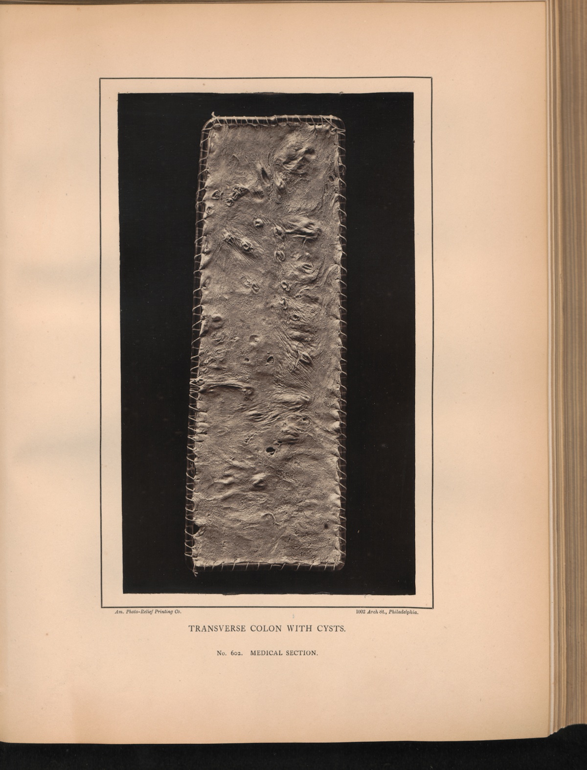 PLATE XXVIII. TRANSVERSE COLON WITH CYSTS. No. 602. MEDICAL
SECTION.
PLATE XXVIII. TRANSVERSE COLON WITH CYSTS. No. 602. MEDICAL
SECTION.
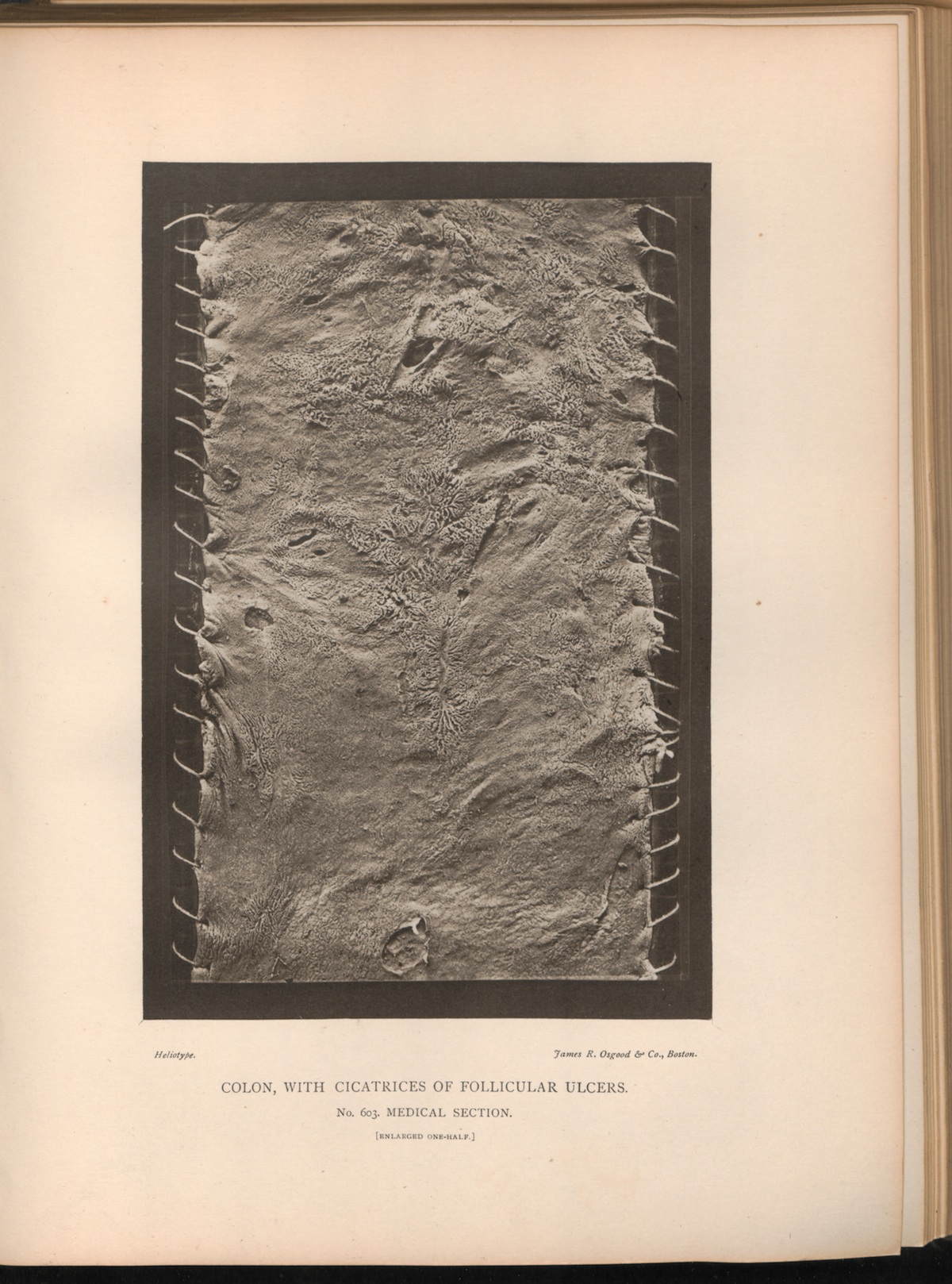 PLATE XXXV. COLON, WITH CICATRICES OF FOLLICULAR ULCERS. No.
603. MEDICAL SECTION. [ENLARGED ONE-HALF.]
PLATE XXXV. COLON, WITH CICATRICES OF FOLLICULAR ULCERS. No.
603. MEDICAL SECTION. [ENLARGED ONE-HALF.]