Title: Smith, Jesse
Source text: Surgeon General Joseph K. Barnes, United States Army, The Medical and Surgical History of the War of the Rebellion. (1861–65.), Part 1, Volume 2 (Washington, D.C.: Government Printing Office, 1870), 59-60.
Civil War Washington ID: med.d1e3676
TEI/XML: med.d1e3676.xml
CASE.—Jesse Smith, Freedman, aged 18 years, employed as a cattle driver, rolled off, in his sleep, from the hay in a stable loft, and fell, some twelve feet to the floor, striking his head. He was found in the morning, cold and insensible, lying on the stable floor, near the horses. Under the use of frictions, hot drinks, and other restoratives he revived, and was carried to the Freedmen's Hospital, at Alexandria, Virginia. Acting Assistant Surgeon Robert N. Atwood, found a wound of the scalp of a crucial form over the right parietal eminence, and a depressed fracture of the bone; but, as the general condition of the patient was comfortable, sensibility being restored, and the mental faculties being apparently normal, Dr. Atwood decided to await further developments. No decidedly bad cerebral symptoms appeared for twelve days after the injury, when the patient complained of increased headache, and a few hours subsequently had a severe convulsion. On the following day, the patient was much the same as usual, except that his headache was increased. Dr. Atwood, in consultation with Acting Assistant Surgeon A. W. K. Andrews, decided to operate, and ether having been administered, enlarged the original wound and applied the trephine, and removed a button of bone to which the greater portion of the depressed fragments were united by the inner table. On removing the bone, pus gushed out copiously. At the upper posterior part of the perforation the inner table was detached three-fourths of an inch more than the outer. This fragment was, with some difficulty, removed by strong forceps. An hour afterwards, the patient having recovered from the ether, was highly excited, restless, and complained of intolerable pain. He was ordered a grain of sulphate of morphia, and in two hours slept comfortably. For ten days subsequently, the morphia was continued, being given to the extent of two or three grains daily. His diet, at this time, was bread and milk, in small quantities, acidulated with vinegar, which he craved earnestly. He also had vinegar and water to drink. In three days after the operation the brain commenced to protrude through the opening in the skull, and by the tenth day had attained the size and shape of half of a hen's egg. Dr. Atwood decided to try, by gentle compression, to reduce the protrusion, and applied a compress and retentive bandage with this view; but immediately violent convulsions occurred; and, although the compress was instantly removed, violent convulsive paroxysms recurred during the night, not less than fifteen or twenty times. The next day the patient was hovering between life and death, but he gradually rallied, and strange to say, after the subsidence of the convulsions he had no more pain in his head. His bowels had been regular throughout his illness, and he had taken no medicine except the morphia, which was discontinued as soon as the pain in the head ceased. Convalescence proceeded rapidly; the protrusion subsided; a firm and dense cicatrix covered the aperture in the skull; and the patient recovered without any impairment of his mental faculties or motor powers. Several months after his recovery he was brought to the Army Medical Museum to be photographed. The picture is numbered 185 in the Surgical Series. The boy was then in perfectly good health, and his faculties were unimpaired. The specimen of the disk and depressed fragment of the parietal was presented to the Museum by Dr. Atwood, and is figured in the accompanying wood cut, (FIG. 25.)
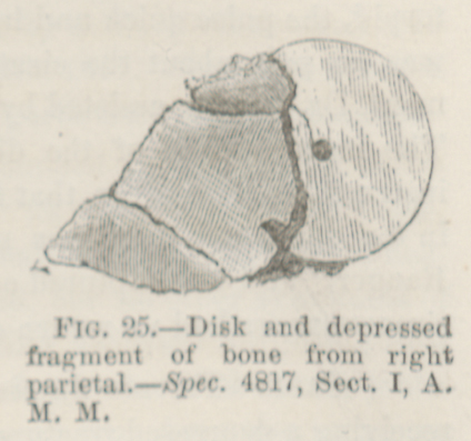 FIG. 25.—Disk and depressed fragment of bone from
right parietal.—Spec. 4817, Sect. I, A. M. M.
FIG. 25.—Disk and depressed fragment of bone from
right parietal.—Spec. 4817, Sect. I, A. M. M.