Title: C——, Thomas P. C.
Source text: Surgeon General Joseph K. Barnes, United States Army, The Medical and Surgical History of the War of the Rebellion. (1861–65.), Part 1, Volume 2 (Washington, D.C.: Government Printing Office, 1870), 568.
Civil War Washington ID: med.d1e20101
TEI/XML: med.d1e20101.xml
CASE.—Private Thomas P. C. C——, Co. A, 9th Mississippi Regiment, aged 19 years, was wounded near Petersburg, Virginia, November 5th, 1864, by a conoidal ball, which entered the posterior portion of the left thorax, passed between the tenth and eleventh ribs, fracturing the latter, as well as the transverse process of the eleventh dorsal vertebra. He was treated in a Confederate hospital at Richmond, and, on May 6th, 1865, was admitted into the hospital at Point Lookout, Maryland. On July 24th, he was transferred to Armory Square Hospital, Washington, and, on August 17th, to Douglas Hospital. The patient stated that on the reception of the injury he had a free hæmorrhage from the wound, spat blood, and had great difficulty of breathing, but that he was doing well until about the middle of June, when he was attacked with diarrhœa. On his admission to Douglas Hospital he was terribly emaciated—almost a living skeleton. On the 18th, the ball, which had lodged behind the tenth rib about one and a half inches from the wound of entrance, was removed by Assistant Surgeon William F. Morris, U. S. A. In spite of the free administration of beef tea and stimulants, with astringents and injections of nitrate of silver for his dysentery, the patient sank rapidly, and died August 20th, 1865. An autopsy was made thirteen hours after death. There were strong pleuritic adhesions between both lungs and the walls of the thorax. The lungs were healthy except the lower lobe of the left lung, which was collapsed and firmly adherent to the walls of the chest near the seat of injury, in such a manner as to form a cavity of considerable dimensions between the wall of the chest and the lung substance. The liver and kidneys were fatty, the spleen healthy, the intestines shrunken and pallid, but everywhere healthy except the descending colon and rectum, where the solitary glands were much enlarged and had ulcerated. They presented the appearance of small cysts, the size of a pea, with minute circular openings at the summit, and contained a transparent gelatinous mass. The pathological specimen is represented in the accompanying wood-cut (FIG. 271). The wound of entrance in the fractured rib is well rounded, and the two are firmly agglutinated by osseous deposit. The preparation was contributed, with a history of the case, by the operator, Assistant Surgeon W. F. Norris, U. S. A.
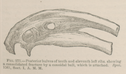 FIG. 271.—Posterior halves of tenth and eleventh left ribs,
showing a consolidated fracture by a conoidal ball, which is attached. Spec. 1561, Sect. I, A. M. M.
FIG. 271.—Posterior halves of tenth and eleventh left ribs,
showing a consolidated fracture by a conoidal ball, which is attached. Spec. 1561, Sect. I, A. M. M.