Title: D——, Thomas
Source text: Surgeon General Joseph K. Barnes, United States Army, The Medical and Surgical History of the War of the Rebellion. (1861–65.), Part 1, Volume 2 (Washington, D.C.: Government Printing Office, 1870), 17-18.
Civil War Washington ID: med.d1e1913
TEI/XML: med.d1e1913.xml
D——, THOMAS, Private, Co. G, 5th Connecticut Volunteers, aged 48 years, was wounded at Chantilly, Virginia, on September 1st, 1862, by several sabre blows over the right ear. He was taken to Washington, and admitted to Douglas Hospital on September 5th. He was then suffering from partial hemiplegia, with mental hebetude. There was great tumefaction of the scalp. It was found that the right parietal was very extensively fractured, (FIG. 2,) one fissure running near the temporo-parietal suture, and others upwards and backwards from the ear. Near the parietal eminence there was a marked depression. It was determined to raise the depressed bone, and on September 6th, Acting Assistant Surgeon J. W. Williams applied the trephine, and, after removing a button and several fragments of bone, he excised a sharp depressed angle by a Hey's saw. It was ascertained that the dura mater had been injured by the sabre-cuts. After the elevation of the depressed fracture, the paralysis of the left side was relieved. The head was shaven, and cold applications were perseveringly employed. For ten days subsequently the case appeared to progress favorably; but, on September 14th, the patient began to be heavy and drowsy, and the following day there were clonic spasms of the left side and pleurosthotonos. At night the breathing was stertorous, the pupils were dilated, and the general symptoms of compression of the brain were very marked. Death took place on September 16th, 1862. There was a large coagulum of extravasated blood under the scalp near the vertex, as though the man had fallen upon his head after being wounded. A post mortem examination was made on September 17th. On removing the calvaria, which was remarkable for its extreme thinness, it was found that the dura mater was perforated beneath the intersection of the wounds, and that, for a space of several inches, there was thickening, with other evidences of inflammatory action. The arachnoid and pia mater were disintegrated in this vicinity, and a space comprising nearly half of the right cerebral hemisphere was occupied by an abscess. The calvaria was forwarded to the Army Medical Museum by Assistant Surgeon Warren Webster, U. S. Army. It is represented by FIG. 2, on the preceding page.
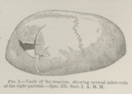 FIG. 2.—Vault of the cranium, showing several
sabre-cuts of the right parietal.—Spec. 235, Sect. I,
A. M. M.
FIG. 2.—Vault of the cranium, showing several
sabre-cuts of the right parietal.—Spec. 235, Sect. I,
A. M. M.