Title: P——, Lyman A.
Source text: Surgeon General Joseph K. Barnes, United States Army, The Medical and Surgical History of the War of the Rebellion. (1861–65.), Part 1, Volume 2 (Washington, D.C.: Government Printing Office, 1870), 393.
Civil War Washington ID: med.d1e18303
TEI/XML: med.d1e18303.xml
CASE.—Sergeant Lyman A. P——, Co. D, 8th Now York Heavy Artillery, aged 21 years, was wounded at Ream's Station, August 25th, 1864, by a musket ball, which entered over the right mastoid process, injured the external ear, and lodged under the skin, a little in front of the auditory foramen. In the difficult retreat, from Ream's Station, he was conveyed to the Base Hospital at City Point, and thence was sent to Washington and admitted to the Lincoln Hospital, on August 28th. The ball had not been extracted, and no symptoms attracted special attention until September 7th, when Acting Assistant Surgeon A. M. Sherman, in charge of the case, observed that the right parotid gland was so greatly inflamed that the patient with difficulty separated his teeth more than one-fourth of an inch. In the course of the day, he had an alarming hæmorrhage, supposed to proceed from the posterior auricular. This was stopped by compression with lint steeped with solution of persulphate of iron. On September 8th, there was no recurrence of bleeding; on the forenoon of the 9th, he had an alarming hæmorrhage, which was temporarily arrested, with difficulty, by compression with lint and styptics, until the patient could be removed to the operating room, when the right primitive carotid was ligated by Acting Assistant Surgeon W. W. Valk, the patient being etherized. The ligature was placed a short distance below the bifurcation, and coagula were removed, and the ball, already mentioned, was extracted from near the angle of the jaw. On the 10th, the patient was quiet, with a frequent pulse; on the 11th, bleeding recurred, and again on the 12th, but ceased spontaneously. There was diarrhœa and vomiting. On the 14th and 15th, the symptoms were regarded by Dr. Sherman as favorable. On the 18th, there were several recurrences of hæmorrhage; on the 19th, there was much swelling of the face and neck, when the ligature was removed. The patient died from hæmorrhage on the following day. The following is an abstract of the notes made at the autopsy, by Acting Assistant Surgeon H. M. Dean: "Height, five feet seven inches. * * * * The submaxillary gland was in a suppurating condition; * * the jugular vein was perfectly normal; there was an abscess extending above and below the point of the artery ligated about three-fourths of an inch; the ligature had come away, and the two extremities of the artery, at the point of ligation, were covered with pus." The wound already adverted to, behind the right ear, was connected with an abscess, which extended down to the angle of the inferior maxilla, and contained a dark-colored fetid pus. The artery from which the hæmorrhage came was not detected. The case is reported by Drs. Sherman and Dean, and Surgeon McKee, in his quarterly report, gives no further remarks on the case by Dr. Valk. The pathological specimen, figured in the wood-cut, shows one-third of the calibre of the vessel undivided. In the wood-cut (FIG. 177), the ligature on the internal carotid was apparently, as Dr. Woodhull has remarked, an experiment upon the cadaver.
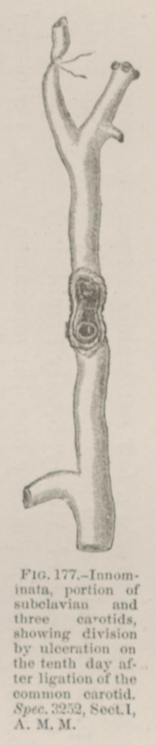 FIG. 177.—Innominata, portion of subclavian and three
carotids, showing division by ulceration on the tenth day after ligation of the common
carotid. Spec. 3252, Sect. I, A. M. M.
FIG. 177.—Innominata, portion of subclavian and three
carotids, showing division by ulceration on the tenth day after ligation of the common
carotid. Spec. 3252, Sect. I, A. M. M.