Title: E——, John
Source text: Surgeon General Joseph K. Barnes, United States Army, The Medical and Surgical History of the War of the Rebellion. (1861–65.), Part 1, Volume 2 (Washington, D.C.: Government Printing Office, 1870), 278.
Civil War Washington ID: med.d1e16914
TEI/XML: med.d1e16914.xml
CASE.—Private John E——, Co. C, 39th New York Volunteers, aged 35 years, was wounded in a melee in a railroad car, near Chicago, Illinois, November 16th, 1862, by a pistol ball, which caused a compound fracture of both tables of the os frontis, right side, near the border of the temporal bone. He arrived at Washington on the 26th, and was admitted to Armory Square Hospital, being quite delirious. There was much tumefaction, and extensive suppuration had already commenced. His strength, appetite, and secretions were normal, his pulse slightly accelerated and tense. An examination revealed a wound of the scalp, two inches in length, crossing the coronal suture at right angles, two inches above the border of the temporal bone. On the morning after admission, the delirium was so intense that it was impossible to keep the patient in the ward. There was occasional difficulty in articulation and a tripping of the left foot in walking, denoting slight hemiplegia. The memory was somewhat impaired, but lucid intervals occurred. The constitutional symptoms had remained the same, denoting irritation rather than compression of the brain. On the 22d, the tumefaction having somewhat subsided, a further examination revealed another wound of the scalp, one and a half inches below the first, and a ball imbedded in the cranium. Ether was administered and an incision made through the integument. The trephine was then applied, and the portion of the cranium containing the ball was removed, as also a fragment of the inner table, triangular in shape, three-eighths of an inch in length and one-fourth of an inch in width at the base. The latter was found driven into the substance of the brain, piercing the dura mater and standing point downward, its broad base displacing the meningea media. After the operation the patient rose from the table, perfectly sane, and walked to his bed. Cold water dressings were applied, laxatives administered, and low diet ordered, and by the 25th he was improving rapidly, there having been no delirium since the operation. On December 10th, the general health of the patient was good, and the wound across the coronal suture had nearly closed by healthy granulations. The wound of operation had healed by first intention, except at the place where the flaps met; at that point there was healthy granulation going on. On January 6th, 1863, he was discharged from the service, having entirely recovered. The specimen removed is a disk of bone, one-half an inch in diameter, with fracture and depression of both tables, and was contributed by Surgeon D. W. Bliss, U. S. V. It is figured in the wood-cut.
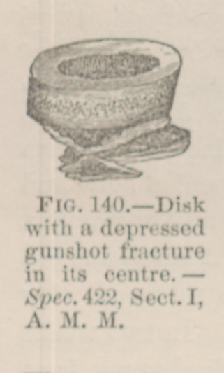 FIG. 140.—Disk with a depressed gunshot fracture its
centre. Spec. 422, Sect. I, A. M. M.
FIG. 140.—Disk with a depressed gunshot fracture its
centre. Spec. 422, Sect. I, A. M. M.